Day: August 13, 2026

  • The Future of Hyperbaric Medicine

    The Future of Hyperbaric Medicine

    How Precision Dosing, Better Trials, Biomarkers, Safety Engineering, and Integrated Care May Shape the Next Era of HBOT

    Hyperbaric medicine is entering a period of greater scientific scrutiny and clinical refinement. The field is moving beyond the question of whether oxygen under pressure produces measurable physiologic effects. Those effects are well established. The more important questions now concern which patients should receive treatment, what oxygen dose is appropriate, how response should be measured, and whether improvement remains meaningful after treatment ends.

    The future of hyperbaric oxygen therapy, commonly abbreviated as HBOT, is unlikely to be defined by simply adding more diagnoses to treatment lists. It will depend on stronger clinical trials, more precise patient selection, standardized outcome reporting, improved safety technology, and closer integration with surgery, wound care, oncology, critical care, and rehabilitation.

    HBOT will continue to have established roles in decompression sickness, arterial gas embolism, carbon monoxide poisoning, delayed radiation injury, selected diabetic foot ulcers, refractory osteomyelitis, acute traumatic ischemia, compromised grafts and flaps, and other recognized conditions. At the same time, neurologic, inflammatory, regenerative, and post-viral applications are being investigated through ongoing trials and research programs. (uhms.org)

    The central challenge will be distinguishing promising investigation from premature clinical adoption.

    Precision Patient Selection Will Become More Important

    Many current hyperbaric indications include clinically diverse patient populations. Two patients may share the same diagnosis while having very different oxygen delivery, vascular reserve, tissue viability, inflammatory activity, and likelihood of responding to treatment.

    A diabetic foot ulcer, for example, may be driven primarily by:

    • Peripheral arterial disease
    • Neuropathy and repetitive pressure
    • Deep infection
    • Osteomyelitis
    • Edema
    • Poor glucose regulation
    • Inadequate debridement
    • A combination of these factors

    HBOT directly influences oxygen delivery, microbial defense, edema, and selected signaling pathways. It cannot independently correct every cause of wound failure.

    Future selection models are likely to combine several types of information:

    • Clinical diagnosis and disease severity
    • Wound or tissue classification
    • Macrovascular blood flow
    • Microvascular oxygen measurements
    • Imaging findings
    • Laboratory and molecular biomarkers
    • Response to initial standard treatment
    • Comorbidities and physiologic reserve
    • Patient-reported symptoms and function

    This approach could reduce treatment of patients whose primary barrier is not oxygen responsive while identifying patients who may benefit before tissue injury becomes irreversible.

    Precision selection will also require clinicians to define the treatment target. A patient may receive HBOT to preserve a flap, stop radiation-related bleeding, reduce tissue necrosis, support infection control, or improve neurologic recovery. Those objectives require different eligibility criteria and different definitions of success.

    HBOT Will Be Treated More Explicitly as a Dose-Dependent Therapy

    A hyperbaric treatment is not defined simply by entering a chamber. The delivered dose depends on several variables:

    • Treatment pressure
    • Inspired oxygen concentration
    • Duration of oxygen exposure
    • Number and length of air breaks
    • Frequency of treatment
    • Number of sessions
    • Time between injury and treatment

    Different combinations may produce different effects on oxygen delivery, edema, inflammation, angiogenic signaling, progenitor-cell mobilization, and oxygen toxicity.

    Historically, many clinical protocols developed through physiology, military experience, expert consensus, and accumulated practice. Future research will increasingly compare different oxygen doses rather than treating all HBOT protocols as equivalent.

    The Hyperbaric Oxygen Brain Injury Treatment Trial, or HOBIT, illustrates this shift. Its adaptive design was developed to evaluate combinations of treatment pressure and oxygen exposure in severe traumatic brain injury and identify the regimen most likely to succeed in a later definitive trial. (clinicaltrials.gov)

    This type of research may help answer questions such as:

    • Is 2.0 ATA sufficient for a particular chronic condition?
    • Does 2.4 or 2.5 ATA improve outcomes enough to justify additional risk?
    • Are two daily treatments necessary during acute tissue salvage?
    • When do air breaks improve safety without reducing efficacy?
    • Can some patients complete fewer sessions without losing benefit?
    • Does a higher dose increase oxygen toxicity without improving the target outcome?

    The future is unlikely to favor one standard protocol for every diagnosis. HBOT may develop more like pharmacology, with indication-specific dosing ranges, response-guided adjustments, and defined stopping criteria.

    Biomarkers May Help Predict Treatment Response

    One of the largest limitations in current practice is the difficulty of predicting response before a lengthy treatment course begins.

    A useful biomarker could help identify whether the biologic pathway targeted by HBOT is active in a particular patient. Potential areas of investigation include:

    • Tissue oxygen response
    • Endothelial function
    • Angiogenic signaling
    • Inflammatory cytokines
    • Oxidative stress markers
    • Circulating endothelial progenitor cells
    • Mitochondrial function
    • Metabolic signatures
    • Fibrosis-associated proteins
    • Genetic or epigenetic factors

    The challenge is not merely finding a laboratory value that changes after treatment. A clinically useful biomarker should predict an outcome that matters, such as wound closure, tissue salvage, reduction in bleeding, avoidance of amputation, improved cognition, or restored function.

    Transcutaneous oxygen measurement already provides a practical example of response-based selection in wound care. Future methods may combine oxygen measurement with perfusion imaging, wound characteristics, and molecular data to create a more reliable probability of benefit.

    Biomarkers could also identify patients at greater risk of adverse effects. Individual variation in oxygen tolerance, seizure threshold, pulmonary response, visual changes, and barotrauma remains incompletely predictable.

    Imaging May Provide Better Evidence of Tissue-Level Change

    Hyperbaric research frequently uses symptom scores, wound measurements, and major clinical outcomes. These remain essential, but advanced imaging may help clarify what changes within the tissue.

    Potential research tools include:

    • Perfusion MRI
    • Functional MRI
    • Diffusion imaging
    • PET imaging
    • Near-infrared spectroscopy
    • Laser Doppler assessment
    • Hyperspectral imaging
    • Microvascular ultrasound
    • Quantitative wound imaging

    In neurologic research, imaging may help identify changes in cerebral perfusion or network activity. In wound and radiation research, imaging may document microvascular recovery, fibrosis, edema, or tissue oxygenation.

    Imaging findings must be interpreted carefully. A change in blood flow or brain activation does not automatically establish improved daily function. Future trials will need to connect imaging biomarkers with patient-centered outcomes rather than treating an imaging change as proof of clinical benefit.

    Better Trial Design Will Shape Which New Indications Survive

    HBOT trials are difficult to design. A credible sham may require placing participants inside a chamber and changing pressure enough to preserve masking. Even modest pressure changes may create physiologic effects, making the comparison treatment imperfect.

    Participants and experienced staff may also recognize differences in pressure, ear sensations, treatment duration, or oxygen-delivery equipment. This can influence expectations and reported symptoms.

    The next generation of trials will increasingly use:

    • Credible sham protocols
    • Blinded outcome assessment
    • Adaptive designs
    • Multicenter enrollment
    • Standardized treatment dosing
    • Clearly defined patient phenotypes
    • Longer follow-up
    • Patient-reported outcomes
    • Health-economic analysis
    • Transparent adverse-event reporting

    An ongoing randomized, blinded, adaptive trial is evaluating HBOT for persistent symptoms after mild to moderate traumatic brain injury in veterans and active-duty service members. Its design reflects the need for stronger masking and more rigorous neurologic outcome measurement. (clinicaltrials.gov)

    Not every emerging indication will survive this level of testing. That is an essential part of scientific progress. A treatment can have a plausible mechanism and produce favorable uncontrolled case series while failing to outperform a credible comparison group.

    Neurologic Applications Will Remain a Major Research Frontier

    The brain is one of the most active areas of hyperbaric investigation. Researchers are studying HBOT in relation to traumatic brain injury, persistent post-concussion symptoms, stroke recovery, cognitive impairment, depression after stroke, and pediatric acquired brain injury.

    Proposed mechanisms include:

    • Increased oxygen availability in metabolically impaired tissue
    • Changes in cerebral blood flow
    • Mitochondrial support
    • Angiogenic signaling
    • Modulation of neuroinflammation
    • Neuroplasticity
    • Recruitment of surviving neural networks

    These mechanisms are scientifically interesting, but neurologic symptoms are complex. Memory, fatigue, pain, sleep, mood, balance, and concentration can change because of rehabilitation, natural recovery, expectation, medication, and concurrent medical care.

    A 2025 systematic review and meta-analysis found continuing interest in HBOT for neurocognitive deficits after traumatic brain injury, but the broader evidence base remains heterogeneous. Ongoing adaptive and placebo-controlled studies are intended to clarify whether specific patient groups and treatment doses produce reproducible functional benefit. (pubmed.ncbi.nlm.nih.gov)

    Neurologic applications are likely to remain investigational until large, well-designed studies demonstrate durable improvements in function rather than isolated changes in imaging or symptom questionnaires.

    Post-Viral and Inflammatory Conditions Will Require Cautious Evaluation

    Long COVID generated substantial interest in HBOT because the condition may involve endothelial dysfunction, autonomic disturbance, inflammation, altered oxygen utilization, fatigue, and cognitive symptoms.

    Early uncontrolled and small controlled studies reported improvements in selected outcomes. More rigorous research has produced a more cautious picture.

    A 2025 randomized, double-blind, placebo-controlled phase 2 trial involving 10 HBOT sessions found that both the HBOT and placebo groups improved, without a significant difference in the primary short-term physical-function outcomes. (pubmed.ncbi.nlm.nih.gov)

    This result does not settle every possible question about long COVID. Different symptom phenotypes, doses, treatment durations, or timing may produce different findings. It does demonstrate that improvement after treatment cannot automatically be attributed to HBOT without an appropriate comparison group.

    Future research will need to define:

    • Which post-viral phenotype is being treated
    • Whether vascular, neurologic, autonomic, or respiratory findings predict response
    • The minimum effective number of sessions
    • Whether improvement persists after treatment
    • How HBOT compares with rehabilitation and other treatments
    • Whether the expected benefit justifies the cost and burden

    Until these questions are answered, post-viral applications should remain within research or carefully governed investigational pathways.

    Delayed Radiation Injury Will Continue to Drive Clinical Research

    Radiation injury is likely to remain one of the strongest areas of HBOT development. Radiation can progressively damage small vessels, reduce tissue oxygenation, increase fibrosis, and impair healing years after cancer treatment.

    The RICH-ART randomized trial demonstrated improvement in patient-reported urinary symptoms following HBOT for chronic radiation cystitis. Five-year follow-up published in 2025 found that clinically meaningful symptom improvement remained durable among many initial responders, although some patients required another course after recurrence. (pubmed.ncbi.nlm.nih.gov)

    Future radiation research will likely focus on:

    • Predicting which patients will respond
    • Comparing pressure and treatment duration
    • Determining the ideal number of sessions
    • Identifying when retreatment is appropriate
    • Measuring fibrosis and vascular recovery
    • Comparing HBOT with endoscopic, pharmaceutical, and surgical alternatives
    • Evaluating cost and treatment burden
    • Studying organ-specific radiation injuries separately

    The evidence will probably continue to become more selective. HBOT may have meaningful value for a defined radiation cystitis population while providing less benefit for another radiation-related symptom complex.

    This distinction is already apparent in head and neck oncology. The 2024 ISOO-MASCC-ASCO guideline found limited evidence supporting routine HBOT for prevention or management of osteoradionecrosis, reinforcing the need to separate established historical practice from current evidence. (pubmed.ncbi.nlm.nih.gov)

    Regenerative Medicine Will Focus on Mechanisms That Translate Into Outcomes

    HBOT influences several processes associated with tissue repair:

    • Angiogenic signaling
    • Fibroblast activity
    • Collagen formation
    • Endothelial function
    • Progenitor-cell mobilization
    • Immune activity
    • Redox-sensitive signaling
    • Mitochondrial metabolism

    These effects have encouraged interest in combining HBOT with regenerative strategies such as:

    • Cellular therapies
    • Tissue-engineered grafts
    • Biomaterial scaffolds
    • Advanced wound matrices
    • Reconstructive surgery
    • Orthobiologic treatments
    • Rehabilitation programs

    The future value of these combinations will depend on more than demonstrating that a marker increased. Researchers will need to show that combination treatment improves graft survival, bone healing, wound closure, function, or another clinically meaningful outcome beyond either treatment alone.

    HBOT may eventually be used to prepare a tissue environment for another intervention or support implanted cells and engineered tissue during the period before a mature blood supply develops. These concepts remain promising but require carefully controlled translational research.

    Cancer Research Will Require Precise Questions

    Questions about HBOT and cancer have historically included concerns that increased oxygen availability might accelerate tumor growth or recurrence. Contemporary reviews have generally not established that appropriately prescribed HBOT promotes malignant progression, but this does not mean it should be used as a cancer treatment outside research.

    The future role of HBOT in oncology is more likely to involve:

    • Management of delayed radiation injury
    • Support for selected reconstructive procedures
    • Treatment of compromised surgical tissue
    • Investigation of oxygen effects on radiation or systemic therapy
    • Reduction of treatment-associated tissue damage

    Any research combining HBOT with active cancer therapy must consider tumor biology, oxygen sensitivity, treatment timing, and possible interactions with chemotherapy, radiotherapy, immunotherapy, and targeted agents.

    Oncologic research should remain tumor specific. Findings involving one cancer type, stage, treatment, or oxygen protocol cannot be generalized across malignancies.

    Real-World Registries Will Complement Randomized Trials

    Rare and emergency hyperbaric conditions are difficult to study through conventional randomized trials. Decompression illness, arterial gas embolism, severe anemia, acute traumatic ischemia, and necrotizing infections may occur too infrequently or require treatment too urgently for easy randomization.

    Multicenter registries can provide valuable information about:

    • Patient characteristics
    • Treatment timing
    • Pressure and oxygen dose
    • Adverse events
    • Clinical outcomes
    • Treatment variation among centers
    • Rare complications
    • Long-term effectiveness

    The UHMS Multicenter Registry for Hyperbaric Oxygen Therapy is designed to collect standardized outcome and safety information for recognized and selected emerging indications. The registry is intended to support research, quality improvement, patient care, and documentation of real-world outcomes. (uhms.org)

    Registry data cannot eliminate selection bias. A patient who receives HBOT may differ from a patient who does not receive it in ways that cannot be fully measured. However, high-quality registry information can help identify treatment patterns, generate hypotheses, evaluate rare outcomes, and guide the design of future trials.

    The field will benefit when participating centers use shared definitions rather than maintaining isolated internal datasets that cannot be compared.

    Safety Engineering Will Become More Visible

    Future progress will depend on safety as much as efficacy.

    Hyperbaric treatment combines elevated pressure, high oxygen exposure, combustible materials, electrical equipment, complex medical devices, and patients who may be critically ill. Fire prevention, maintenance, product review, grounding, staff training, and emergency preparedness are fundamental clinical requirements.

    In August 2025, the FDA reminded healthcare providers to follow chamber instructions for use, maintain strict fire-prevention controls, monitor patients continuously, conduct required maintenance, and control electrical or static-producing items. The communication followed reports of serious injuries and deaths associated with HBOT devices. (fda.gov)

    Future chamber and facility development may include:

    • More advanced oxygen monitoring
    • Automated detection of environmental abnormalities
    • Improved grounding and static-control systems
    • Better equipment compatibility testing
    • Integrated treatment verification
    • More reliable gas-source identification
    • Improved critical-care monitoring under pressure
    • Enhanced maintenance analytics
    • Standardized emergency alerts
    • Better human-factor design

    Automation may reduce certain errors, but it will not replace qualified staff. A system can detect an abnormal pressure or oxygen value, but trained personnel must understand its clinical meaning and respond safely.

    Data Systems May Reduce Treatment Errors

    Hyperbaric programs generate detailed treatment information, including pressure profiles, oxygen periods, air breaks, patient observations, glucose values, equipment checks, and adverse events.

    Improved digital integration could allow:

    • Automatic transfer of treatment profiles into the medical record
    • Verification of the prescribed protocol against the delivered protocol
    • Alerts for cumulative oxygen exposure
    • Tracking of adverse-event patterns
    • Identification of missed reassessment intervals
    • Comparison of outcomes across indications
    • Maintenance and equipment alerts
    • More complete accreditation documentation

    Clinical decision support might eventually identify a mismatch between a patient’s diagnosis, prescribed pressure, payer requirements, and continued-treatment criteria.

    These systems must be designed carefully. Poorly configured alerts can create fatigue, while automated documentation can reproduce incorrect information at scale. Technology should support clinical judgment and safety checks rather than replace them.

    Artificial Intelligence May Assist With Selection and Monitoring

    Artificial intelligence could eventually combine clinical, imaging, oxygenation, wound, and laboratory data to estimate the likelihood of treatment response.

    Potential applications include:

    • Predicting wound-healing probability
    • Identifying patients at risk of treatment failure
    • Quantifying wound area and tissue characteristics from photographs
    • Detecting changes in graft or flap viability
    • Recognizing unusual treatment-monitoring patterns
    • Forecasting missed appointments or course noncompletion
    • Supporting utilization review

    These applications remain early. Any predictive model would require diverse, high-quality datasets and external validation across different hospitals, chambers, patient populations, and payer environments.

    An algorithm trained primarily on one type of wound or one health system may perform poorly elsewhere. Clinical teams will also need to understand which variables drive a recommendation and whether those variables reflect genuine biology or historical referral patterns.

    AI should not become a method of expanding treatment volume without clinical justification.

    Critical-Care Hyperbaric Medicine May Become More Regionalized

    Some hyperbaric indications involve unstable patients who need mechanical ventilation, vasoactive medication, invasive monitoring, surgery, or intensive care. These include arterial gas embolism, severe carbon monoxide poisoning, necrotizing infection, acute traumatic ischemia, and exceptional blood-loss anemia.

    Not every hyperbaric center can or should manage this level of acuity.

    The future may involve stronger regional networks in which:

    • Community hospitals identify indications early
    • Hyperbaric physicians provide rapid consultation
    • Transfer criteria are standardized
    • Critical-care capable chambers receive complex patients
    • Transport teams understand pressure-related emergencies
    • Referring hospitals continue stabilization while transfer is arranged

    Teleconsultation may improve early decision-making, particularly for diving emergencies, carbon monoxide poisoning, radiation complications, and potential graft or flap compromise.

    Remote consultation cannot replace physical examination or chamber staffing. Its value is in helping the referring team decide whether transfer is justified and what care should continue before transport.

    Workforce Development Will Be Essential

    Expanding clinical capability requires trained physicians, nurses, technologists, respiratory therapists, safety leaders, and engineers.

    Future workforce development will need to address:

    • Formal hyperbaric education
    • Chamber-specific competency
    • Physician credentialing and privileging
    • Critical-care training
    • Emergency simulation
    • Fire-safety education
    • Continuing professional development
    • Succession planning
    • Coverage for after-hours emergencies

    Programs that depend on one physician, one technologist, or one safety specialist are operationally fragile. Staff retirement, illness, or turnover can reduce capability immediately.

    The future of the specialty will depend on creating durable professional pathways rather than treating hyperbaric medicine as a secondary responsibility added to an unrelated clinical role.

    Accreditation and Standardization Will Become More Important

    As HBOT expands into different hospital, outpatient, and private settings, variation in equipment, staffing, training, and clinical governance becomes more visible.

    Accreditation provides a structure for evaluating:

    • Facility safety
    • Medical leadership
    • Staff qualifications
    • Treatment protocols
    • Patient selection
    • Equipment maintenance
    • Emergency readiness
    • Documentation
    • Quality improvement
    • Clinical outcomes

    The UHMS accreditation program evaluates the facility as an integrated clinical system rather than assessing the chamber alone. (uhms.org)

    Patients, referring clinicians, hospitals, and payers may increasingly expect evidence that a center follows recognized standards. Accreditation cannot guarantee a favorable outcome, but it can reduce reliance on informal practices and individual memory.

    Economic Research Will Influence Clinical Adoption

    Many HBOT courses require daily treatment over several weeks. The financial cost includes more than chamber time. Patients may face transportation, missed work, caregiving requirements, copayments, and treatment fatigue.

    Hospitals must account for:

    • Chamber acquisition and construction
    • Oxygen and compressed-air systems
    • Staffing
    • Maintenance
    • Fire protection
    • Accreditation
    • Physician supervision
    • Prior authorization
    • Treatment cancellations
    • Compliance monitoring

    Future studies will increasingly evaluate cost per meaningful clinical outcome rather than cost per chamber session.

    Relevant outcomes may include:

    • Amputations avoided
    • Operations avoided
    • Hospital admissions prevented
    • Transfusions reduced
    • Wound-care duration shortened
    • Function restored
    • Quality-adjusted life years
    • Return to work
    • Reduced recurrence

    A treatment may be expensive per session but cost effective if it prevents a major amputation or complex reconstruction. Another treatment may improve a short-term symptom score without producing enough durable benefit to justify a lengthy course.

    Economic evaluation should not be used merely to reduce access. It should help direct HBOT toward patients most likely to receive meaningful value.

    Treatment Access Will Remain Uneven

    Access to medical HBOT varies substantially by geography. Some regions have hospital-based programs capable of treating ventilated patients and diving emergencies. Others have outpatient wound-focused centers but no critical-care chamber. Rural or coastal areas may be many hours from recompression capability.

    Improving access may require:

    • Regional transfer agreements
    • Shared on-call coverage
    • Better emergency referral protocols
    • Mobile consultation systems
    • Transportation assistance
    • More efficient outpatient scheduling
    • Strategic placement of critical-care chambers

    Increasing the number of chambers is not enough. A chamber without qualified staff, maintenance, safety infrastructure, and medical governance does not create reliable access.

    Access also includes the ability to complete treatment. A patient who cannot travel five days per week may receive no practical benefit from a theoretically appropriate prescription.

    Low-Pressure Wellness Services Will Remain Separate From Medical HBOT

    Commercial interest in low-pressure chambers, wellness oxygen services, recovery programs, and anti-aging treatment is likely to continue.

    These services should not be confused with hospital-grade HBOT delivered for a defined medical indication. Differences may include:

    • Chamber pressure
    • Oxygen concentration
    • Device clearance
    • Fire protection
    • Medical supervision
    • Staff competency
    • Emergency capability
    • Treatment objective
    • Supporting evidence

    A 2025 systematic review found that evidence supporting HBOT for aesthetic medicine and anti-aging remained limited and did not justify broad clinical claims. (pubmed.ncbi.nlm.nih.gov)

    The future credibility of hyperbaric medicine will depend partly on maintaining this distinction. Expanding unsupported claims may create public interest in the short term, but it can weaken scientific trust, increase safety risk, and make evidence-based clinical programs harder to distinguish.

    What a Future-Ready Hyperbaric Program Will Look Like

    A mature hyperbaric program will not be defined primarily by the number of chambers it operates.

    It will demonstrate:

    • Clearly defined clinical indications
    • Qualified medical and safety leadership
    • Diagnosis-specific treatment protocols
    • Objective response criteria
    • Multidisciplinary care pathways
    • Strong fire-prevention systems
    • Chamber-compatible critical-care capability when offered
    • Structured adverse-event review
    • Participation in accreditation and registries
    • Transparent clinical outcomes
    • Ethical management of emerging indications
    • Financial and documentation compliance

    The program will also be willing to stop treatment when the objective has been reached or when evidence shows that continued exposure is unlikely to help.

    This discipline is essential. A predetermined course should not continue automatically because authorization was obtained or chamber capacity is available.

    The Most Important Research Priorities

    Several priorities are likely to determine how the specialty develops:

    1. Identifying patient phenotypes most likely to respond.
    2. Comparing different pressures and oxygen doses.
    3. Developing credible sham protocols.
    4. Linking biomarkers and imaging to clinical outcomes.
    5. Standardizing outcomes across centers.
    6. Measuring durability after treatment.
    7. Evaluating treatment burden and cost effectiveness.
    8. Improving safety and device surveillance.
    9. Building multicenter registries for rare indications.
    10. Separating therapeutic evidence from promotional claims.

    The next era of hyperbaric medicine will be defined less by whether HBOT can influence biology and more by whether clinicians can apply that biology predictably.

    The field has compelling physiologic mechanisms, established emergency applications, and growing evidence for selected chronic tissue injuries. It also has areas where enthusiasm continues to exceed certainty.

    Progress will require both openness and restraint. Researchers must remain willing to investigate new applications, while clinicians must be willing to describe those applications as investigational until credible evidence demonstrates meaningful benefit.

    The future of hyperbaric medicine is therefore not unlimited expansion. It is greater precision, safer delivery, stronger evidence, and better integration with the treatments that patients already need.

  • HBOT in Burn Treatment

    HBOT in Burn Treatment

    The Clinical Role of Hyperbaric Oxygen in Acute Thermal Injury, Smoke Exposure, Tissue Preservation, and Burn Reconstruction

    Severe burns are dynamic injuries. Tissue damage can continue after the heat source has been removed because edema, microvascular thrombosis, inflammation, and impaired oxygen delivery may convert marginally viable tissue into deeper necrosis.
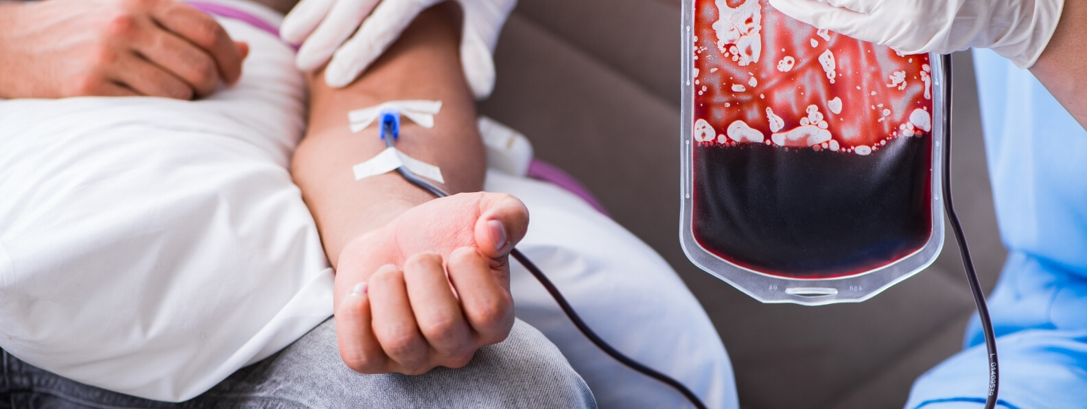
    Hyperbaric oxygen therapy, commonly abbreviated as HBOT, may be considered as an adjunct for selected acute thermal burns. Its proposed role is to increase oxygen delivery to threatened tissue, reduce edema, support microvascular function, and potentially limit the progression of burn depth.

    The Undersea and Hyperbaric Medical Society recognizes acute thermal burn injury as a clinical indication for adjunctive HBOT. The available evidence, however, does not support routine chamber treatment for every burn. HBOT should be delivered only when it can be integrated safely with burn-center care and without delaying airway management, fluid resuscitation, wound excision, grafting, or another essential intervention. (UHMS)

    Burn Depth and Tissue Viability

    Burn severity is determined by more than the percentage of skin involved. Clinicians must also consider depth, location, inhalation injury, associated trauma, age, comorbidities, and the patient’s physiologic response.

    Burns are generally classified as:

    • Superficial: Injury limited to the epidermis
    • Superficial partial thickness: Injury extending into the upper dermis
    • Deep partial thickness: More extensive dermal injury with reduced blanching and a greater risk of scarring or grafting
    • Full thickness: Destruction of the entire dermis
    • Deep tissue injury: Extension into fat, fascia, muscle, or bone

    Superficial burns are not included in calculations of total body surface area. Partial-thickness and full-thickness burns are included. The American Burn Association recommends immediate burn-center consultation for full-thickness burns, partial-thickness burns involving at least 10 percent of total body surface area, and deep burns involving the face, hands, feet, genitalia, perineum, or major joints. (American Burn Association)

    A thermal burn is often described as having three tissue zones. The central zone of coagulation contains irreversibly injured tissue. The surrounding zone of stasis has impaired circulation but may remain viable. The outer zone of hyperemia generally has better perfusion and a stronger likelihood of recovery.

    The zone of stasis is the principal potential target of adjunctive HBOT. Treatment cannot restore tissue that has already undergone irreversible coagulative necrosis. Its proposed benefit is preservation of hypoxic but viable tissue surrounding the deepest portion of the burn.

    Why Burn Wounds Can Become Deeper

    A burn that initially appears partial thickness can progress into a deeper injury during the first several days. This process is commonly called burn wound conversion.

    Factors contributing to conversion may include:

    • Microvascular thrombosis
    • Endothelial injury
    • Capillary leakage
    • Progressive edema
    • Inflammatory cell activation
    • Vasoconstriction
    • Infection
    • Hypotension or inadequate resuscitation
    • Repeated pressure or trauma
    • Drying or inappropriate wound management

    The microcirculation may be particularly compromised during the first 12 to 24 hours, while the injury can remain biologically active for approximately 72 hours. This provides the rationale for beginning adjunctive HBOT early when it is selected. (NCBI)

    Preventing burn conversion can have important clinical consequences. Preserving even a portion of the dermis may reduce the area requiring excision, improve epithelial regeneration, decrease grafting requirements, and limit long-term contracture or hypertrophic scarring.

    HBOT should not be expected to prevent conversion when perfusion has been lost completely, shock remains uncontrolled, or tissue is already necrotic.

    Standard Burn Care Remains the Foundation

    Initial burn management follows established trauma and critical-care priorities. These should never be postponed for chamber treatment.

    Depending on the injury, essential care may include:

    • Airway evaluation and early intubation
    • High-concentration oxygen
    • Hemodynamic stabilization
    • Intravenous fluid resuscitation
    • Burn-depth and surface-area assessment
    • Escharotomy or fasciotomy
    • Wound cleansing and debridement
    • Early excision of deep burns
    • Skin grafting or flap reconstruction
    • Temperature control
    • Pain and anxiety management
    • Nutritional support
    • Infection surveillance
    • Physical and occupational therapy

    Suspected inhalation injury requires burn-center consultation. The American Burn Association recommends immediate consultation with consideration for transfer for all patients with suspected inhalation injury. (American Burn Association)

    HBOT should be viewed as a supporting intervention within this system. A patient who needs airway control, escharotomy, hemorrhage management, or surgery should receive that intervention first.

    How HBOT Changes Oxygen Delivery

    During HBOT, the patient breathes oxygen while exposed to an ambient pressure higher than normal atmospheric pressure. The elevated pressure substantially increases arterial oxygen tension and the amount of oxygen dissolved directly in plasma.

    This may allow oxygen-rich plasma to move through functioning microvessels and diffuse farther into tissue surrounding the burn. Potentially relevant effects include:

    • Increased oxygen delivery to hypoxic but viable tissue
    • Reduced edema through hyperoxic vasoconstriction
    • Preservation of marginal microcirculation
    • Support for cellular energy production
    • Reduced leukocyte adhesion during reperfusion injury
    • Improved oxygen-dependent microbial killing
    • Support for fibroblast and epithelial activity
    • Promotion of vascular repair

    The combination of increased plasma oxygen and controlled vasoconstriction is important. Vasoconstriction may reduce capillary pressure and fluid leakage while the elevated oxygen content helps maintain tissue oxygen delivery.

    HBOT does not restore circulation through a completely thrombosed artery, correct inadequate fluid resuscitation, or remove devitalized tissue. Enough functioning circulation must remain for oxygenated plasma to reach the wound.

    Edema Reduction in Acute Burns

    Edema is a major component of burn pathophysiology. Thermal injury increases capillary permeability, allowing fluid and proteins to move into the interstitial space. Large burns can produce both local swelling and systemic intravascular volume depletion.

    Within the wound, edema increases the distance oxygen must diffuse between capillaries and cells. Swelling may also compress small vessels and worsen microvascular perfusion.

    HBOT may reduce edema by producing vasoconstriction in oxygenated tissue while preserving oxygen delivery through the increased plasma oxygen concentration. Experimental and clinical reports have suggested that this may help interrupt the cycle of edema, ischemia, and additional capillary injury. (UHMS)

    This effect does not replace appropriate burn resuscitation. Large burns may require substantial fluid administration, and the patient’s urine output, hemodynamics, lactate, electrolytes, and organ function must continue to be monitored.

    Attempts to reduce edema by withholding necessary resuscitation can worsen tissue ischemia and organ injury.

    HBOT and Burn Wound Conversion

    One proposed benefit of early HBOT is the preservation of the zone of stasis. Increased oxygen delivery may support cells that are metabolically stressed but not yet irreversibly injured.

    The treatment may also influence secondary injury by reducing leukocyte-endothelial adhesion, inflammatory signaling, and ischemia-reperfusion effects. These mechanisms could help limit the progression of a deep partial-thickness wound into a full-thickness injury.

    Clinically meaningful preservation would be reflected by:

    • Less progression of burn depth
    • More spontaneous epithelialization
    • Smaller areas requiring excision
    • Reduced grafting requirements
    • Shorter time to wound closure
    • Improved tissue quality

    Temporary redness or improved color during chamber exposure does not establish tissue salvage. The benefit must persist outside the chamber and translate into durable wound healing.

    Which Burn Patients May Be Considered for HBOT?

    There is no universally accepted set of burn-specific selection criteria. Practice varies by burn center, hyperbaric facility, injury severity, and chamber availability.

    A consultation may be considered for selected patients with:

    • Extensive deep partial-thickness burns

    • Burns at substantial risk of wound conversion

    • Severe edema threatening tissue viability

    • Circumferential burns after appropriate decompression

    • Burns involving critical functional areas

    • High-voltage electrical injuries with traumatic ischemia

    • Compromised grafts or flaps after burn reconstruction

    • Carbon monoxide poisoning associated with a fire

    • Burn injury combined with another recognized hyperbaric indication

    Factors making HBOT less appropriate may include:

    • Superficial or uncomplicated burns

    • Completely necrotic tissue requiring excision

    • Uncontrolled shock

    • An untreated pneumothorax

    • An airway that cannot be managed safely in the chamber

    • Immediate need for surgery

    • Lack of chamber-compatible critical-care equipment

    • Transport that would disrupt burn-center treatment

    The most useful question is not simply whether the patient has a burn. It is whether HBOT can plausibly preserve viable tissue or treat a separate recognized complication without compromising standard care.

    Treatment Timing

    If HBOT is selected for an acute thermal burn, early initiation is generally favored because the zone of stasis is most salvageable before secondary microvascular failure becomes established.

    Published protocols commonly describe beginning treatment during the first 24 hours when possible. Some regimens use several treatments during the initial day, followed by once- or twice-daily sessions during the early period of burn progression. Reported treatment pressures often range from approximately 2.0 to 2.4 atmospheres absolute, with oxygen exposure lasting around 90 minutes. (NCBI)

    These values should not be interpreted as a universal prescription. The 2026 systematic review of current burn evidence found substantial variation in pressure, frequency, treatment duration, number of sessions, patient severity, and reported outcomes. (PubMed Central (PMC))

    The hyperbaric physician and burn surgeon should determine:

    • Whether the injury remains salvageable
    • How urgently treatment should begin
    • How chamber sessions will fit around operations
    • Whether once- or twice-daily treatment is feasible
    • Which clinical findings justify continuation
    • When HBOT should be discontinued

    A necessary operation always takes priority over a scheduled chamber session.

    Smoke Inhalation and Airway Injury

    Smoke inhalation can produce several distinct problems:

    • Upper-airway thermal injury
    • Chemical irritation of the tracheobronchial tree
    • Pulmonary inflammation and edema
    • Carbon monoxide poisoning
    • Cyanide poisoning
    • Acute respiratory failure

    HBOT does not directly remove airway casts, reverse upper-airway edema, or replace mechanical ventilation. Patients may require early intubation, bronchoscopy, pulmonary hygiene, ventilator support, and burn-intensive-care management. (PubMed)

    The role of HBOT is clearer when the fire exposure has caused clinically significant carbon monoxide poisoning. Carbon monoxide reduces oxygen transport and can produce neurologic and cardiac injury. HBOT may be considered according to the patient’s neurologic findings, loss of consciousness, cardiac involvement, acidosis, pregnancy status, exposure severity, and transport logistics.

    Suspected cyanide poisoning should be treated promptly with appropriate antidotal and critical care, commonly including hydroxocobalamin when clinically indicated. Chamber treatment must not delay antidote administration or airway stabilization. (PubMed)

    Smoke inhalation should therefore not be treated as one uniform hyperbaric diagnosis. The clinician must distinguish airway injury, pulmonary damage, carbon monoxide toxicity, cyanide toxicity, and the cutaneous burn itself.

    Burn Surgery, Grafting, and Reconstruction

    Deep partial-thickness and full-thickness burns frequently require excision and grafting. HBOT should not delay removal of nonviable eschar or definitive wound coverage.

    It may become relevant when:

    • The recipient bed is poorly oxygenated
    • A graft is failing to establish adequate uptake
    • A flap develops ischemia or venous congestion
    • Radiation, trauma, or infection has compromised the wound bed
    • Marginal tissue around a reconstruction remains salvageable

    The role in a compromised graft or flap is clinically distinct from routine treatment of an acute burn. Medicare recognizes preparation and preservation of compromised skin grafts as a covered indication, although it does not cover HBOT for the primary management of ordinary wounds. (Centers for Medicare & Medicaid Services)

    A threatened graft or flap first requires surgical evaluation. Hematoma, seroma, pressure, infection, pedicle kinking, thrombosis, and other correctable causes must be addressed. HBOT may then support viable hypoxic tissue while vascular connections recover or mature.

    Infection and Immune Function

    Loss of the skin barrier, devitalized tissue, invasive devices, prolonged hospitalization, and immune dysfunction all contribute to infection risk after major burns.

    HBOT can increase oxygen-dependent microbial killing by neutrophils and may improve oxygen delivery to tissue receiving antimicrobial therapy. It may also support granulation and wound defense in selected hypoxic regions.

    It cannot replace:

    • Surgical excision of infected or necrotic tissue
    • Drainage of abscesses
    • Culture-directed antimicrobial therapy
    • Sepsis management
    • Removal of infected devices
    • Definitive wound closure

    The 2026 systematic review identified signals suggesting improved infection control in some studies, but variability in study design and outcomes prevented definitive conclusions. (PubMed Central (PMC))

    HBOT should not be continued simply because a burn is infected. The team must identify whether a recognized oxygen-responsive problem remains after source control has been addressed.

    What Current Evidence Shows

    The clinical evidence for HBOT in burn treatment is mixed.

    A 2026 systematic review included 13 studies, consisting of five randomized controlled trials, seven cohort studies, and one case-control study. The studies evaluated 566 burn patients treated with HBOT and varied considerably in burn severity, treatment protocols, and outcome measures. The review found promising signals related to healing, infection, progression of burn depth, surgery, and hospital stay, but it could not establish definitive effects on mortality or long-term outcomes. (PubMed Central (PMC))

    Earlier controlled research produced inconsistent results. A randomized prospective study in a referral burn-center population did not demonstrate a significant overall benefit from HBOT. (PubMed)

    A Cochrane review concluded that there was insufficient evidence to support or refute routine HBOT for thermal burns. The small trials were clinically heterogeneous and had substantial methodologic limitations. A separate trial involving burn wounds treated with split-thickness grafting reported greater graft survival, but the result was not enough to justify routine treatment of all acute burns. (PubMed)

    The evidence can therefore be summarized cautiously:

    • HBOT has a credible physiologic rationale.
    • Some studies report improved wound healing and reduced complications.
    • Other controlled research has found little or no benefit.
    • Patient selection and treatment protocols are not standardized.
    • High-quality evidence for mortality reduction is lacking.
    • Routine treatment of every burn is not supported.

    This uncertainty makes multidisciplinary selection more important, not less.

    Measuring Whether HBOT Is Helping

    The treatment team should define objective goals before beginning HBOT.

    Potential measures include:

    • Change in burn depth
    • Area of preserved dermis
    • Reduction in edema
    • Rate of epithelialization
    • Area requiring excision
    • Graft or flap viability
    • Number of operative procedures
    • Time to wound closure
    • Infection status
    • Functional tissue preservation
    • Length of hospitalization

    Serial photography, standardized wound assessment, operative findings, perfusion testing, and graft-survival measurements may help document response.

    Treatment should be reconsidered when:

    • Necrosis continues to progress
    • A surgical problem remains uncorrected
    • The patient becomes unstable
    • Treatment repeatedly disrupts burn care
    • No measurable clinical benefit is developing
    • The original treatment objective has been achieved

    A predetermined number of sessions should not replace ongoing clinical judgment.

    Treating Critically Ill Burn Patients Under Pressure

    Major burn patients may require mechanical ventilation, continuous infusions, invasive monitoring, large-volume resuscitation, temperature control, and frequent surgical intervention.

    HBOT should be provided only in a facility capable of preserving that level of care under pressure. Operational requirements may include:

    • Chamber-compatible ventilators
    • Infusion pumps able to function under pressure
    • Secure airway and vascular access
    • Continuous physiologic monitoring
    • Management of chest tubes and drains
    • Temperature maintenance
    • Safe wound dressings
    • Staff experienced in hyperbaric critical care
    • Rapid access to emergency decompression

    Transporting the patient from the burn intensive care unit to the chamber can itself create risk. Lines may become displaced, temperature may fall, infusions may be interrupted, and access to the patient may be limited during treatment.

    The potential tissue benefit must be substantial enough to justify these operational burdens.

    Fire Safety and Burn Dressings

    The hyperbaric environment requires strict control of combustible materials, ignition sources, electrical equipment, clothing, linens, and wound products.

    Burn patients may arrive with topical preparations, antimicrobial creams, petroleum-containing products, synthetic dressings, warming equipment, or electronic devices that have not been approved for chamber use. Every item must undergo a documented hyperbaric safety review.

    The FDA advises facilities to follow the chamber manufacturer’s instructions, maintain continuous patient monitoring, use appropriate grounding, perform scheduled maintenance, and enforce fire-prevention procedures. (U.S. Food and Drug Administration)

    A dressing that is appropriate in the burn unit is not automatically appropriate inside a pressurized oxygen environment. The burn, hyperbaric, pharmacy, and safety teams should agree on compatible alternatives before treatment begins.

    Risks of HBOT in Burn Patients

    Potential complications include:

    • Middle-ear or sinus barotrauma
    • Pulmonary barotrauma
    • Oxygen-induced seizure
    • Temporary vision changes
    • Claustrophobia or agitation
    • Glucose instability
    • Interruption of critical-care therapies
    • Hypothermia during transport
    • Fire-safety hazards
    • Delayed access during sudden deterioration

    An untreated pneumothorax must be addressed before pressurization. This is especially important after blast injury, inhalation injury, chest trauma, or mechanical ventilation.

    Patients with facial burns, airway edema, sedation, or altered mental status may be unable to report ear pain or neurologic symptoms. The chamber team must anticipate complications rather than depending solely on patient communication.

    Reimbursement and Coverage Considerations

    Professional recognition and insurance coverage are not the same.

    Although UHMS recognizes acute thermal burns as a hyperbaric indication, the current Medicare National Coverage Determination lists thermal skin burns as nationally noncovered. Medicare also excludes acute thermal or chemical pulmonary damage described as smoke inhalation with pulmonary insufficiency. (Centers for Medicare & Medicaid Services)

    Separate complications may qualify under another covered indication. Medicare covers conditions including acute carbon monoxide intoxication, cyanide poisoning, acute traumatic peripheral ischemia, crush injury, and preparation or preservation of compromised skin grafts. (Centers for Medicare & Medicaid Services)

    Commercial payer policies vary. Before nonemergency treatment, the hospital should verify:

    • The diagnosis being submitted
    • Whether the indication is covered
    • Prior-authorization requirements
    • Required clinical documentation
    • Treatment limits
    • Facility and professional billing requirements

    Coverage should not determine emergency stabilization, but it affects program planning and informed discussions about treatment burden.

    Integrating HBOT Into Burn-Center Care

    An effective hospital pathway should define:

    1. Which burn injuries prompt urgent hyperbaric consultation.
    2. Which burn surgeon remains responsible for definitive wound management.
    3. How airway, resuscitation, and operative priorities are protected.
    4. Which patients can be monitored safely inside the available chamber.
    5. How HBOT is coordinated with excision, grafting, and dressing changes.
    6. Which objective findings justify continued treatment.
    7. How adverse events and outcomes are reviewed.

    The decision should be made jointly by burn surgery, critical care, hyperbaric medicine, respiratory therapy, nursing, pharmacy, and hyperbaric safety personnel.

    HBOT in burn treatment is best understood as an early tissue-preservation strategy for carefully selected patients. It may increase oxygen delivery, reduce edema, protect marginal microcirculation, and support graft or flap survival. It does not replace resuscitation, airway management, surgery, infection control, nutrition, or rehabilitation.

    Current evidence is promising but inconsistent. The most responsible use is selective, protocol driven, and tied to measurable clinical goals within a specialized burn-care system.

  • HBOT for Severe Anemia

    HBOT for Severe Anemia

    Using Hyperbaric Oxygen as a Temporary Bridge When Red Blood Cell Transfusion Is Unavailable or Declined

    Severe anemia reduces the blood’s capacity to transport oxygen to the brain, heart, kidneys, and other metabolically active tissues. When oxygen delivery falls below cellular demand, the patient may develop myocardial ischemia, neurologic dysfunction, lactic acidosis, organ failure, or death.

    Red blood cell transfusion is ordinarily the fastest and most effective way to restore oxygen-carrying capacity in life-threatening anemia. Hyperbaric oxygen therapy, commonly abbreviated as HBOT, may be considered when transfusion is not possible because compatible blood cannot be obtained, the patient declines blood products, severe hemolysis prevents effective transfusion, or an extraordinary blood shortage limits access.

    The Undersea and Hyperbaric Medical Society recognizes severe anemia as a clinical indication for HBOT when transfusion cannot be performed. In this setting, HBOT is not intended to correct the anemia itself. It temporarily increases the amount of oxygen dissolved in plasma while bleeding is controlled and the patient produces new red blood cells. (UHMS)

    What Severe Anemia Means in Hyperbaric Medicine

    The relevant hyperbaric indication is sometimes described as severe anemia or exceptional blood-loss anemia. It does not refer to every patient with a low hemoglobin level.

    Routine iron-deficiency anemia, mild postoperative anemia, anemia of chronic disease, and stable nutritional anemia are generally treated by identifying the cause and restoring iron, vitamin, renal, marrow, or hormonal function. HBOT is reserved for exceptional circumstances in which oxygen delivery has become critically inadequate and red blood cell transfusion cannot be used promptly.

    Potential scenarios include:

    • Massive obstetric, gastrointestinal, traumatic, or surgical hemorrhage
    • Severe anemia in a patient who declines transfusion
    • Multiple red blood cell antibodies that make compatible blood difficult to obtain
    • Autoimmune hemolysis with ongoing red blood cell destruction
    • Delayed access to blood during a disaster or remote emergency
    • A severe transfusion reaction that temporarily prevents additional transfusion
    • Rare situations in which blood products are medically contraindicated

    Current AABB guidance recommends considering transfusion at a hemoglobin concentration below approximately 7 g/dL for many hemodynamically stable hospitalized adults. That general threshold should not be confused with the much more critical condition in which HBOT may be considered. Transfusion decisions must account for active bleeding, symptoms, hemodynamics, cardiac disease, organ dysfunction, and the overall clinical context. (DOI)

    Recent UHMS literature describes HBOT as a bridge for patients with severe anemia when transfusion is impossible because of religious objection, crossmatch incompatibility, or blood unavailability. The decision should be driven by evidence of inadequate tissue oxygen delivery rather than a hemoglobin number in isolation. (UHMS)

    Why a Normal Pulse Oximeter Can Be Misleading

    Most oxygen carried in blood is bound to hemoglobin. A much smaller quantity is dissolved directly in plasma.

    A conventional pulse oximeter estimates the percentage of available hemoglobin binding sites occupied by oxygen. It does not measure how much hemoglobin is present. A patient with critically low hemoglobin can therefore display an oxygen saturation of 100 percent while having severely inadequate total oxygen content.

    Clinical evidence of oxygen debt may include:

    • Persistent tachycardia
    • Hypotension
    • Chest pain or ischemic electrocardiographic changes
    • Altered mental status
    • Syncope
    • Dyspnea
    • Rising lactate
    • Metabolic acidosis
    • Reduced urine output
    • Myocardial injury
    • Progressive organ dysfunction

    The relationship between hemoglobin concentration and clinical instability is not identical for every patient. A gradual decline may permit physiologic adaptation, while rapid blood loss can produce collapse at a higher measured hemoglobin. Fever, pain, sepsis, pregnancy, cardiac disease, and increased metabolic demand can further reduce tolerance.

    How HBOT Increases Oxygen Delivery

    During HBOT, the patient breathes oxygen while exposed to increased atmospheric pressure. This produces a marked increase in arterial oxygen tension and in the quantity of oxygen physically dissolved in plasma.

    At sufficiently high treatment pressures, dissolved plasma oxygen can temporarily provide a substantial portion of resting tissue oxygen requirements even when hemoglobin is critically limited. Plasma can also move through functioning microvessels that may be difficult for red blood cells to traverse during low-flow or edematous states. (UHMS)

    This physiologic effect is immediate but temporary. Oxygen delivery rises while the patient is breathing oxygen under pressure and then declines after decompression.

    HBOT does not:

    • Replace lost red blood cells
    • Stop active hemorrhage
    • Correct iron deficiency
    • Reverse bone marrow failure
    • Eliminate hemolysis
    • Permanently increase hemoglobin after one treatment

    Its purpose is to relieve accumulating oxygen debt while definitive treatment takes effect. Studies evaluating blood counts have not shown HBOT itself to produce an immediate increase in hemoglobin, hematocrit, or red blood cell count. (PubMed)

    HBOT Functions as a Bridge, Not a Transfusion Substitute

    Red blood cell production requires time. Even after erythropoietin, iron, folate, vitamin B12, and other deficiencies are addressed, the marrow cannot immediately replace a major loss of circulating red cells.

    HBOT may create repeated periods of enhanced tissue oxygenation during this vulnerable interval. Treatments can be scheduled according to the return of clinical signs of oxygen debt, with the interval between sessions lengthened as hemoglobin rises and organ function stabilizes.

    This pulsed strategy is conceptually different from a standard weekday wound-care course. Severe anemia may require urgent, prolonged, or repeated treatments within the first several days, accompanied by intensive care monitoring. UHMS guidance describes treatment pressures generally ranging from 2.0 to 3.0 atmospheres absolute, with air breaks and treatment duration adjusted to the patient’s clinical condition and recurrence of oxygen debt. (UHMS)

    HBOT should be discontinued when the patient can maintain adequate oxygen delivery outside the chamber, transfusion becomes possible, or the risks of continued treatment exceed the expected benefit.

    Controlling Blood Loss Is the First Priority

    HBOT cannot compensate indefinitely for ongoing hemorrhage. The source of blood loss must be identified and controlled as quickly as possible.

    Depending on the cause, this may require:

    • Emergency surgery
    • Endoscopic hemostasis
    • Interventional radiology and embolization
    • Obstetric hemorrhage control
    • Reversal of anticoagulation
    • Antifibrinolytic medication
    • Vascular repair
    • Direct pressure, packing, or topical hemostatic agents

    The treatment team should also determine which blood components, fractions, medications, and procedures the patient is willing or able to receive. A patient who declines red blood cell transfusion may accept some clotting factors, albumin, cell salvage, erythropoietin, or other therapies. Preferences vary and should never be assumed from religious identity alone. (AAFP)

    For a patient with decision-making capacity, an informed refusal of transfusion should be respected and documented. The discussion should include the risk of irreversible organ injury or death, available alternatives, and the limitations of HBOT.

    A Comprehensive Blood-Conservation Plan

    HBOT should be one component of a broader patient blood management strategy.

    The plan may include:

    • Immediate control of bleeding
    • High-concentration oxygen between chamber treatments
    • Intravenous iron when iron availability is inadequate
    • Erythropoiesis-stimulating medication when clinically appropriate
    • Folate and vitamin B12 replacement
    • Correction of vitamin or nutritional deficiencies
    • Optimization of ventilation and cardiac output
    • Treatment of fever, pain, agitation, and shivering
    • Reduction of unnecessary metabolic demand
    • Limitation of diagnostic blood draws
    • Use of low-volume laboratory tubes
    • Point-of-care testing
    • Avoidance of unnecessary intravenous fluid dilution

    Minimizing phlebotomy is especially important. Repeated laboratory testing can remove a clinically meaningful amount of blood from a patient who has almost no red cell reserve. Testing should be consolidated, performed only when it will alter management, and completed with the smallest practical sample volume. (AAFP)

    Iron and erythropoiesis-stimulating agents should not be presented as immediate oxygen-delivery treatments. Their purpose is to accelerate red blood cell recovery over subsequent days. Adequate iron availability is necessary for an effective erythropoietic response.

    Investigational Oxygen-Carrying Products

    Hemoglobin-based oxygen carriers have occasionally been used as emergency bridges when red blood cell transfusion is impossible. These products are designed to transport oxygen without intact donor red blood cells.

    They are not routinely approved for general clinical use in the United States. Access may require emergency regulatory authorization, and potential adverse effects include vasoconstriction, hypertension, thrombosis, myocardial injury, and increased mortality. (AAFP)

    Published cases have combined HBOT with an investigational hemoglobin-based oxygen carrier, iron, erythropoietin, hemostatic treatment, and intensive care support. These reports demonstrate what may be possible in exceptional circumstances, but they do not establish a standard protocol suitable for every hospital. (PubMed)

    Hyperbaric Treatment Protocols for Severe Anemia

    There is no single HBOT schedule appropriate for every patient with life-threatening anemia.

    The prescription may account for:

    • Hemoglobin concentration and rate of decline
    • Whether bleeding has stopped
    • Lactate and acid-base status
    • Neurologic condition
    • Cardiac ischemia
    • Hemodynamic support
    • Mechanical ventilation
    • Oxygen requirements outside the chamber
    • Response to previous HBOT sessions
    • Evidence that erythropoiesis has begun
    • Treatment-related adverse effects

    UHMS describes initial treatment pressures in the range of approximately 2.0 to 3.0 ATA. Oxygen may be delivered in intervals separated by air breaks, and an initial session may continue for several hours in an unstable patient. Repeated treatment is then guided by the return of symptoms or laboratory evidence of oxygen debt. (UHMS)

    This differs from routine elective HBOT protocols. A patient with exceptional anemia may require treatment outside normal operating hours, close coordination with the intensive care unit, and repeated physician reassessment.

    Treatment should occur only in a hospital-based hyperbaric facility capable of managing the patient’s full acuity.

    Treating a Critically Ill Patient Under Pressure

    Patients with life-threatening anemia may require:

    • Mechanical ventilation
    • Continuous electrocardiographic monitoring
    • Vasopressor or inotropic medication
    • Invasive arterial monitoring
    • Active temperature control
    • Sedation
    • Postoperative drains
    • Ongoing hemostatic treatment

    The chamber team must be able to maintain these interventions safely under pressure. Ventilators, infusion devices, monitoring equipment, vascular lines, and airway systems must be evaluated for hyperbaric compatibility.

    A multiplace chamber may allow an attendant to remain with the patient, but it still requires trained inside and outside personnel. A monoplace chamber may accommodate a ventilated patient in selected facilities, but access during treatment is limited.

    The decision to initiate HBOT should consider whether transporting the patient from the operating room or intensive care unit creates an unacceptable risk. The chamber should support critical care rather than interrupt it.

    Monitoring for Recurrent Oxygen Debt

    Hemoglobin concentration remains important, but treatment decisions should incorporate the complete physiologic picture.

    Monitoring may include:

    • Mental status
    • Heart rate and blood pressure
    • Electrocardiographic changes
    • Cardiac biomarkers
    • Serum lactate
    • Blood gas analysis
    • Acid-base status
    • Urine output
    • Renal and hepatic function
    • Peripheral perfusion
    • Oxygen requirements
    • Hemoglobin and reticulocyte trends

    A falling lactate, improved cognition, resolution of ischemic changes, and stable organ function outside the chamber may support longer intervals between treatments. Recurrent acidosis, chest pain, confusion, hypotension, or organ dysfunction may indicate renewed oxygen debt.

    No single hemoglobin concentration guarantees safety. The patient’s oxygen demand, cardiovascular reserve, rate of blood loss, and response to treatment all influence tolerance.

    Risks of HBOT in Severe Anemia

    Hyperbaric treatment has recognized risks, including:

    • Middle-ear or sinus barotrauma
    • Pulmonary barotrauma
    • Oxygen-induced seizure
    • Temporary visual changes
    • Claustrophobia
    • Hemodynamic instability during transport
    • Disruption of intensive care
    • Complications involving chamber-incompatible equipment

    An untreated pneumothorax must be corrected before pressurization. Severe pulmonary disease, unstable cardiac conditions, seizure risk, and implanted devices require individualized evaluation.

    Oxygen toxicity deserves particular attention because severe anemia protocols may involve relatively high pressures, prolonged exposure, or repeated sessions. Scheduled air breaks help limit uninterrupted oxygen exposure, but continuous clinical observation remains essential.

    The practical risks may be greater than the direct chamber risks. Transporting an unstable patient away from the operating room, blood bank, or intensive care team can delay urgent intervention. HBOT is appropriate only when the facility can preserve the required level of care throughout preparation, treatment, and decompression.

    Clinical Evidence Supporting HBOT

    The evidence for HBOT in severe anemia differs from the randomized trial evidence available for more common medical treatments.

    A systematic review evaluated 35 publications describing the use of HBOT for severe anemia. The reports generally described favorable clinical outcomes, but the literature consisted largely of case reports, case series, physiologic studies, and expert experience rather than randomized controlled trials. (PubMed)

    Randomized trials are difficult because the treatment is used in rare, life-threatening situations where transfusion is impossible. Patients also receive multiple simultaneous interventions, making it difficult to isolate the contribution of HBOT.

    One frequently cited case involved a patient with massive obstetric hemorrhage whose hemoglobin fell to 2.0 g/dL. Management included critical care, ventilatory support, erythropoietin, and pulsed HBOT. The report demonstrates the bridge concept, but a single successful case cannot establish the expected outcome for other patients. (PubMed)

    A 2021 case report similarly described HBOT as part of a bloodless management strategy for severe pernicious anemia. The case broadens the clinical experience but does not mean HBOT should routinely be used for vitamin B12 deficiency or other chronic anemias. (NCBI)

    The most accurate interpretation is that HBOT has a strong physiologic rationale and supportive clinical experience as a rescue bridge, while high-certainty comparative evidence remains limited.

    HBOT Is Not Routine Treatment for Chronic Anemia

    Patients with stable anemia generally need evaluation and treatment of the underlying cause.

    That may include:

    • Oral or intravenous iron
    • Vitamin B12 or folate
    • Treatment of gastrointestinal or menstrual blood loss
    • Management of kidney disease
    • Treatment of inflammation or malignancy
    • Modification of marrow-suppressive medication
    • Hematology evaluation
    • Red blood cell transfusion when clinically indicated

    HBOT should not be marketed as a method for increasing routine energy levels, treating low ferritin, improving athletic performance, or correcting uncomplicated anemia.

    A patient with iron deficiency needs iron and evaluation of the source of deficiency. A patient with marrow failure needs hematologic treatment. A patient with active bleeding needs hemostasis. Hyperbaric oxygen becomes relevant only when tissue oxygen delivery is critically threatened and ordinary restoration of red blood cell capacity is not immediately possible.

    Coverage and Reimbursement Limitations

    Clinical recognition does not guarantee insurance coverage.

    Although UHMS recognizes severe anemia as a hyperbaric indication, the current Medicare National Coverage Determination explicitly lists exceptional blood-loss anemia as a nationally noncovered condition. (Centers for Medicare & Medicaid Services)

    Commercial payer policies may differ, but emergency authorization and reimbursement can be challenging. Hospitals considering this service should address:

    • Payer authorization
    • Emergency financial approval
    • Medical-necessity documentation
    • Physician and facility billing
    • Uncompensated emergency treatment
    • Transfer agreements with regional centers

    Coverage limitations should be discussed early, but financial questions should not delay clinically necessary emergency stabilization. The facility’s compliance, finance, and case-management teams may need to work alongside critical care and hyperbaric clinicians.

    Multidisciplinary Management Is Essential

    Severe anemia requiring HBOT is not solely a hyperbaric problem. It requires coordination among:

    • Critical care
    • Hematology
    • Surgery or the specialty controlling the bleeding
    • Anesthesiology
    • Transfusion medicine
    • Pharmacy
    • Laboratory medicine
    • Ethics and legal services
    • Patient blood management
    • Hyperbaric medicine

    The team should establish a shared plan for controlling hemorrhage, minimizing additional blood loss, supporting erythropoiesis, monitoring organ function, and determining when HBOT can be reduced or discontinued.

    For patients who decline transfusion, communication should remain respectful and precise. Declining one treatment does not mean declining all medical care. The team should clarify acceptable interventions privately, assess decision-making capacity, document the patient’s choices, and avoid making assumptions about which products or procedures the patient will accept. (AAFP)

    HBOT for severe anemia is best understood as temporary physiologic support during an extraordinary emergency. It raises dissolved plasma oxygen while clinicians stop blood loss and rebuild red blood cell mass. It does not cure anemia, and it does not provide the sustained oxygen-carrying capacity of circulating red blood cells.

    When transfusion is impossible and tissue oxygen debt is progressing, however, appropriately delivered HBOT may provide the time needed for definitive treatment and endogenous blood recovery to succeed.

  • HBOT for Compromised Skin Grafts

    HBOT for Compromised Skin Grafts

    Using Hyperbaric Oxygen Therapy to Support Graft Uptake, Preserve Viable Tissue, and Reduce the Need for Repeat Reconstruction

    Skin grafting allows surgeons to cover wounds that cannot be closed primarily. Grafts are used after trauma, burns, tumor removal, infection control, debridement, and reconstructive procedures. Successful grafting can protect exposed tissue, reduce fluid loss, improve function, and shorten the time required for wound closure.
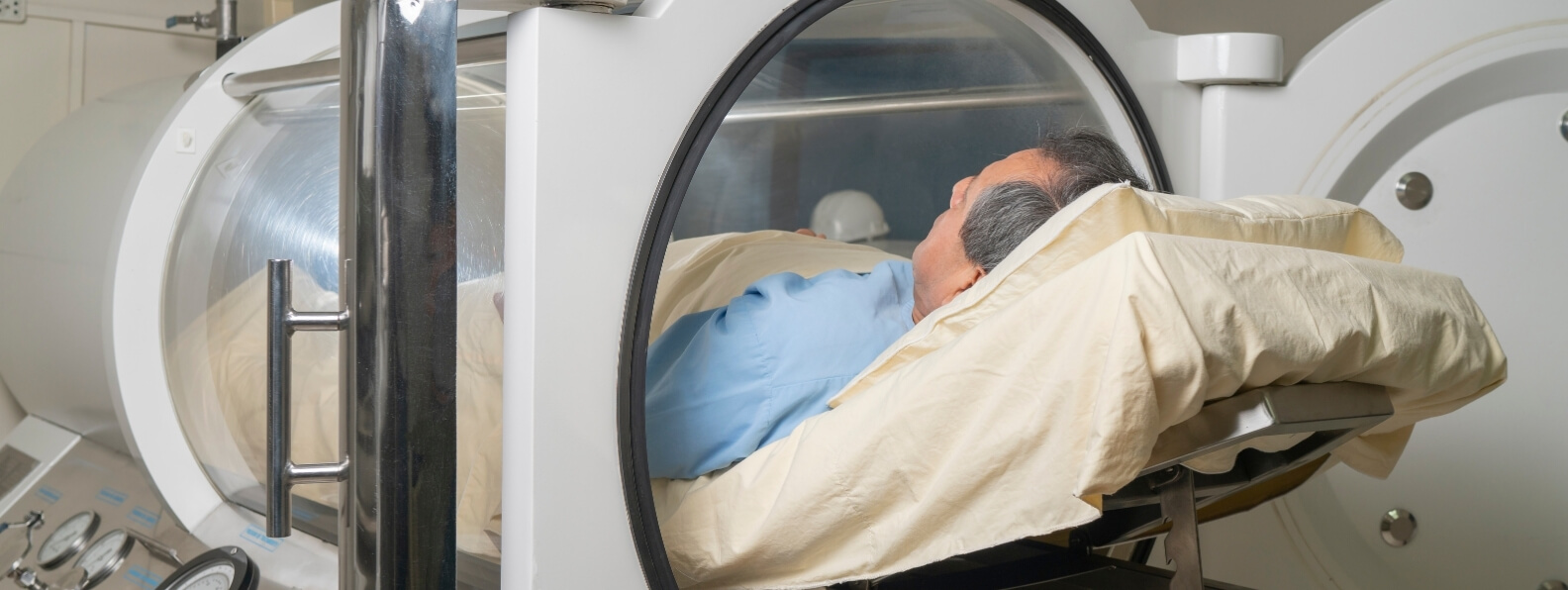
    Unlike a flap, a skin graft is completely separated from its original blood supply. It must survive temporarily through diffusion from the recipient bed and then establish new vascular connections. Any process that interferes with contact, oxygen delivery, or revascularization can cause partial or complete graft loss.

    Hyperbaric oxygen therapy, commonly abbreviated as HBOT, may be considered when a skin graft becomes compromised by hypoxia or impaired perfusion. It is not recommended routinely for a healthy graft that is progressing normally. The Undersea and Hyperbaric Medical Society identifies compromised grafts and flaps as an accepted indication, emphasizing that HBOT is a salvage adjunct for threatened tissue rather than routine support for uncomplicated reconstruction. (UHMS)

    How a Skin Graft Establishes a Blood Supply

    A skin graft does not immediately receive blood through its own circulation after placement. Early survival depends on a staged relationship with the recipient wound bed.

    During the first phase, known as plasmatic imbibition, the graft absorbs oxygen, nutrients, and fluid from the underlying tissue. This diffusion-based support helps preserve graft cells while more permanent vascular connections are developing.

    During inosculation, small vessels within the graft align with vessels in the recipient bed. Blood flow begins to enter the graft as these vascular channels connect.

    Revascularization then progresses through capillary ingrowth and remodeling. The graft gradually develops a more dependable circulation capable of supporting long-term survival.

    These stages require:

    • Close contact between the graft and wound bed
    • An adequately vascularized recipient surface
    • Control of bleeding and fluid accumulation
    • Protection from movement and shear
    • Management of infection
    • Sufficient tissue oxygenation

    A disruption during the early postoperative period may prevent vascular connection and lead to graft separation, ischemia, or necrosis.

    Split-Thickness and Full-Thickness Skin Grafts

    A split-thickness skin graft contains the epidermis and a portion of the dermis. Because it is relatively thin, it can receive nutrients by diffusion more readily and may survive on a less vascular recipient bed than a thicker graft.

    Split-thickness grafts are commonly used to cover:

    • Large traumatic wounds
    • Burn wounds
    • Chronic ulcers
    • Donor sites
    • Wounds following debridement
    • Areas prepared with dermal substitutes

    A full-thickness skin graft contains the epidermis and the complete dermis. It generally provides better durability, contour, texture, and resistance to contraction, but it has greater metabolic requirements and depends on a well-vascularized recipient bed.

    HBOT may be relevant to either graft type when tissue oxygenation is inadequate. The graft’s thickness, location, recipient bed, wound cause, and degree of compromise all influence the likelihood of salvage.

    Why Skin Grafts Become Compromised

    A compromised graft is one that is at meaningful risk of partial or complete failure. The underlying cause must be identified quickly because HBOT cannot correct every mechanism of graft loss.

    Common causes include:

    • Hematoma beneath the graft
    • Seroma or fluid accumulation
    • Infection
    • Shear or excessive movement
    • Poor graft fixation
    • Inadequate recipient-bed preparation
    • Arterial insufficiency
    • Venous congestion
    • Severe edema
    • Previous radiation exposure
    • Diabetes-related microvascular dysfunction
    • Tobacco or nicotine exposure
    • Excessive pressure from dressings
    • Repeated trauma
    • Malnutrition
    • Immunosuppression
    • Extensive crush or burn injury

    Exposed cortical bone without periosteum, tendon without paratenon, cartilage without perichondrium, and poorly vascularized scar tissue may not provide an adequate recipient bed unless additional preparation or reconstruction is performed.

    HBOT should not be used to avoid correcting a mechanical, vascular, infectious, or surgical cause of graft failure.

    Recognizing Early Graft Compromise

    Early recognition is important because viable but hypoxic tissue may still be salvageable. Once graft tissue becomes irreversibly necrotic, oxygen therapy cannot restore it.

    Potential warning findings include:

    • Increasing pallor
    • Dusky, blue, or violaceous discoloration
    • Persistent coolness
    • Delayed capillary refill
    • Progressive edema
    • Blistering or epidermal separation
    • Loss of adherence to the wound bed
    • Fluid collecting beneath the graft
    • Malodor or purulent drainage
    • Darkening tissue
    • Failure to demonstrate expected vascularization
    • Expanding areas of necrosis

    Color alone is not always reliable. A graft may appear pale during the earliest phase before vascular flow is fully established. A congested graft may appear dark even though some tissue remains viable. Clinical interpretation should consider timing, temperature, adherence, bleeding characteristics, drainage, infection, and the condition of the wound bed.

    Serial photographs and consistent documentation can help distinguish normal postoperative evolution from progressive compromise.

    Surgical Evaluation Comes Before HBOT

    A threatened graft requires prompt evaluation by the surgeon or reconstructive team. Correctable causes should be addressed immediately.

    Potential interventions include:

    • Evacuating a hematoma
    • Draining a seroma
    • Removing a constricting dressing
    • Reapplying an appropriate bolster
    • Correcting graft displacement
    • Debriding devitalized tissue
    • Treating infection
    • Improving arterial inflow
    • Relieving venous congestion
    • Revising the wound bed
    • Returning the patient to the operating room

    A graft that is floating on blood or fluid cannot reliably establish vascular contact. HBOT may increase oxygen availability, but it cannot press the graft back against the recipient bed.

    Similarly, HBOT cannot overcome an untreated arterial occlusion or a wound bed that is structurally incapable of supporting graft uptake. The treatment becomes most relevant after remediable causes have been corrected and viable tissue remains at risk.

    How HBOT May Support a Compromised Graft

    During HBOT, the patient breathes oxygen while exposed to increased atmospheric pressure. The treatment raises arterial oxygen tension and substantially increases the amount of oxygen dissolved directly in plasma.

    This oxygen-rich plasma can move through functioning vessels in the wound bed and create a stronger diffusion gradient into the graft. That effect may be especially useful during the early period when the graft has not yet developed an independent circulation.

    Potential benefits include:

    • Increased oxygen diffusion from the recipient bed
    • Support for cellular metabolism during plasmatic imbibition
    • Improved fibroblast activity
    • Support for collagen formation
    • Enhanced capillary ingrowth
    • Promotion of angiogenic signaling
    • Reduction of edema through hyperoxic vasoconstriction
    • Improved oxygen-dependent leukocyte microbial killing
    • Modulation of ischemia-reperfusion injury

    Reviews of compromised grafts and flaps describe increased tissue oxygenation, neovascularization, fibroblast support, and reduction of ischemia-reperfusion injury as important mechanisms through which HBOT may improve salvage. (PubMed Central (PMC))

    These effects depend on some remaining perfusion. HBOT cannot deliver a clinically meaningful oxygen dose into tissue that has no functioning vascular access.

    Oxygen Diffusion During the Early Graft Period

    The temporary lack of direct circulation makes a newly placed graft particularly dependent on diffusion.

    Under ordinary atmospheric conditions, oxygen delivery into a hypoxic wound bed may be inadequate because of edema, microvascular injury, radiation damage, or arterial disease. Increasing dissolved plasma oxygen strengthens the gradient between the wound bed and the graft.

    This may help maintain marginal cells until vascular connections mature. It may also support the recipient bed, where endothelial cells, fibroblasts, leukocytes, and extracellular matrix must work together to secure and vascularize the graft.

    HBOT does not make the graft independent of its wound bed. It temporarily improves the oxygen environment in which graft adherence and revascularization must occur.

    Edema Control and Graft Adherence

    Edema can compromise graft survival by increasing diffusion distance and reducing microvascular blood flow. Swelling may also contribute to mechanical separation between the graft and recipient surface.

    Hyperoxic vasoconstriction can reduce blood flow volume in selected tissues while the elevated oxygen content of plasma helps preserve oxygen delivery. This combination may reduce edema without creating the degree of tissue hypoxia that ordinary vasoconstriction might produce.

    The practical objective is not merely to make the wound look less swollen. It is to improve the relationship between capillaries, the recipient bed, and the graft.

    Edema control should still include appropriate elevation, dressing selection, treatment of venous obstruction, and correction of any constricting or mechanically disruptive factor.

    HBOT and Infection Risk

    Bacterial contamination and infection can interfere with graft adherence, damage the recipient bed, and increase local oxygen demand.

    HBOT may support oxygen-dependent microbial killing by neutrophils and enhance the activity of selected antimicrobial agents. These effects may improve the local defense environment, but HBOT is not a replacement for infection management.

    Treatment may still require:

    • Drainage
    • Debridement
    • Deep tissue cultures
    • Culture-directed antibiotics
    • Removal of infected material
    • Management of systemic sepsis
    • Delayed regrafting after infection control

    A graft covering an inadequately controlled infection is unlikely to survive solely because tissue oxygenation has increased.

    Previous Radiation Increases Graft Risk

    Radiated tissue may become fibrotic, hypovascular, and chronically hypoxic. Small-vessel damage can progress for years after radiation treatment, leaving the wound bed with limited capacity to support graft uptake.

    When graft compromise occurs within a radiation field, the patient may have overlapping hyperbaric indications:

    • Preservation of a compromised graft
    • Treatment of soft tissue radionecrosis
    • Treatment of osteoradionecrosis when bone is involved

    The clinical team should also evaluate for recurrent malignancy, infection, exposed bone, fistula, and other causes of tissue breakdown.

    HBOT may improve oxygenation and stimulate vascular remodeling in viable irradiated tissue, but it does not remove necrotic tissue or eliminate the need for appropriate oncologic and reconstructive evaluation.

    When to Begin HBOT

    Timing matters. HBOT is most biologically plausible while graft tissue is hypoxic but still viable.

    The hyperbaric team should be contacted when compromise is recognized, not only after the graft has become black, dry, and clearly necrotic. Delaying consultation may reduce the amount of tissue that can be preserved.

    A practical sequence is:

    1. Identify signs of graft compromise.
    2. Notify the operating or reconstructive surgeon.
    3. Correct hematoma, seroma, pressure, displacement, infection, or vascular obstruction.
    4. Determine whether viable tissue remains.
    5. Begin HBOT promptly when the graft is still considered salvageable.
    6. Reassess the graft and wound bed throughout treatment.
    7. Return to surgery if progressive necrosis or another correctable problem develops.

    The chamber schedule should accommodate surgical care. A necessary operation should not be postponed to complete an HBOT session.

    Selecting Patients for Hyperbaric Treatment

    HBOT may be considered when:

    • A graft is clinically compromised rather than merely at theoretical risk
    • Hypoxia or impaired perfusion is contributing to failure
    • Mechanical causes have been corrected
    • The recipient bed retains some blood supply
    • The graft contains tissue that remains potentially viable
    • Salvage would reduce wound size, repeat surgery, or reconstructive complexity
    • The patient can be treated safely under pressure
    • The surgical and hyperbaric teams can coordinate frequent reassessment

    HBOT is less likely to provide meaningful benefit when:

    • The graft is healing normally
    • The graft is already completely necrotic
    • A hematoma or seroma remains untreated
    • The graft is detached from the wound bed
    • A major arterial obstruction has not been corrected
    • An uncontrolled infection requires surgery
    • The recipient bed cannot support vascular ingrowth
    • Treatment would delay a more urgent intervention

    The decision should be based on whether HBOT can plausibly change the clinical outcome, not simply whether a skin graft is present.

    HBOT Treatment Protocols

    Protocols vary according to the type and severity of compromise, timing, wound cause, and facility practice.

    Commonly described regimens use:

    • Approximately 2.0 to 2.5 atmospheres absolute
    • About 90 to 120 minutes of oxygen exposure
    • One or two treatments daily during the initial salvage period
    • Transition to once-daily treatment as viability stabilizes
    • Continued reassessment rather than a fixed course applied to every patient

    Reviews based on UHMS practice recommendations commonly describe twice-daily treatment initially, followed by daily treatment when the graft or flap demonstrates improved viability. The total course depends on the clinical response and may be shorter than courses used for chronic radiation injury or diabetic wounds. (PubMed Central (PMC))

    The treatment prescription should be individualized by a qualified hyperbaric physician. More pressure or more treatments do not automatically create better graft survival.

    Monitoring Graft Response

    The treatment team should define objective salvage goals before beginning HBOT.

    Serial assessment may include:

    • Graft color
    • Temperature
    • Capillary refill
    • Adherence to the wound bed
    • Percentage of viable graft
    • Drainage
    • Infection findings
    • Edema
    • Demarcation of necrotic areas
    • Need for additional debridement
    • Progress toward stable wound coverage

    Photographic documentation can help quantify changes over time. The surgeon should remain involved because the significance of color, adherence, and tissue loss depends on the graft type, recipient site, operation, and expected reconstructive outcome.

    Meaningful benefit may include:

    • Increased percentage of graft uptake
    • Prevention of further graft loss
    • Reduction in the area requiring regrafting
    • Preservation of coverage over exposed structures
    • Avoidance of a more complex flap procedure
    • Earlier progression to stable wound closure

    A graft should not be described as salvaged merely because it appears temporarily pinker during oxygen exposure. The improvement should persist outside the chamber and translate into durable tissue survival.

    Evidence Supporting HBOT

    The evidence base includes animal studies, physiologic research, clinical series, reviews, and a limited number of controlled trials.

    A 2017 review concluded that HBOT may increase composite graft survival, improve skin graft outcomes, and support compromised flap salvage. The authors also emphasized the importance of correcting mechanical causes and initiating treatment promptly after compromise is recognized. (PubMed Central (PMC))

    A 2020 UHMS review similarly described supportive basic science and clinical evidence across several graft and flap types. However, much of the clinical literature consists of case reports, case series, and nonrandomized studies, which are vulnerable to selection bias and inconsistent outcome reporting. (PubMed)

    A Cochrane review of HBOT for acute surgical and traumatic wounds found that the available trials were small and at risk of bias. One study suggested improved split-skin graft survival in burn wounds, while other studies did not establish consistent benefit. The review concluded that high-quality evidence was insufficient for broad routine use. (PubMed Central (PMC))

    More recently, a 2025 randomized trial involving 64 adults undergoing split-thickness grafting for traumatic wounds reported higher mean graft uptake with HBOT on postoperative days 4 and 7. Mean graft uptake on day 7 was approximately 91.7 percent in the HBOT group and 83.1 percent in the standard-care group. Donor-site healing was also faster in the HBOT group. (PubMed)

    That trial strengthens the evidence that HBOT can influence graft uptake in selected traumatic wounds. It does not prove that every uncomplicated graft should receive routine treatment, nor does it directly answer every question about rescuing a graft after compromise has already developed. The population, wound cause, treatment protocol, and resources available must be considered before generalizing the results.

    Routine Prophylactic HBOT Is Not the Standard

    UHMS guidance states that HBOT is neither necessary nor recommended for normal, uncompromised grafts and flaps. (UHMS)

    A newly placed graft may be clinically important without being compromised. Routine postoperative HBOT would expose many patients to treatment burden and risk without clear evidence of added benefit.

    Selective preoperative or immediate postoperative treatment may sometimes be considered when the recipient bed is predictably high risk, such as severely irradiated tissue or a reconstruction with very limited options. In these circumstances, the indication should be documented precisely. The rationale is tissue hypoxia or a recognized radiation injury, not simply the presence of a graft.

    The 2025 randomized trial may encourage further research into planned perioperative treatment for high-risk traumatic grafts. It should not be used to justify automatic HBOT after every split-thickness graft. (PubMed)

    Medicare Coverage and Medical Necessity

    Medicare’s national coverage determination includes the preparation and preservation of compromised skin grafts. It specifically clarifies that HBOT is not covered under this indication for the primary management of wounds. (Centers for Medicare & Medicaid Services)

    Documentation should therefore establish:

    • That a graft is present or being prepared in a clinically appropriate setting
    • Why the graft is compromised
    • The evidence of hypoxia, ischemia, or impaired viability
    • Corrective surgical measures already performed
    • The specific objective of HBOT
    • Serial evidence of response
    • Continued coordination with the reconstructive team

    Payer policies differ. Prior authorization, diagnosis coding, treatment limits, and documentation requirements should be verified for each patient.

    Coverage should not determine clinical judgment, but weak documentation can create both reimbursement and compliance risk.

    Standard Graft Care Must Continue

    HBOT supports the wound environment but does not replace meticulous graft management.

    The care plan may still include:

    • Appropriate graft fixation
    • Bolster or negative-pressure therapy
    • Control of fluid beneath the graft
    • Immobilization
    • Pressure protection
    • Limb elevation
    • Infection management
    • Vascular assessment
    • Glucose management
    • Nutritional support
    • Tobacco and nicotine cessation
    • Timely dressing changes
    • Surgical debridement

    Negative-pressure wound therapy may improve graft contact, remove fluid, and reduce shear in selected wounds. HBOT and negative-pressure therapy address different barriers and may be used together when clinically appropriate.

    Nutrition is also important. A patient with inadequate protein, calories, vitamins, or trace elements may have impaired collagen production, immune activity, and epithelial repair despite improved oxygenation.

    Tobacco and Nicotine Exposure

    Tobacco smoke exposes tissue to carbon monoxide and other substances that interfere with oxygen transport and vascular function. Nicotine can produce vasoconstriction and may further compromise blood flow to a vulnerable wound bed.

    Patients should receive clear cessation counseling rather than being told simply that smoking is discouraged. Continued exposure may undermine both the graft and the physiologic objective of HBOT.

    The hyperbaric team should also follow strict facility policies regarding smoking, ignition sources, clothing, dressings, and skin products. The FDA advises hyperbaric facilities to follow device instructions, maintain fire-prevention systems, train staff, monitor patients continuously, and control materials introduced into the chamber environment. (U.S. Food and Drug Administration)

    Risks of HBOT

    HBOT is generally well tolerated when delivered in an appropriately staffed medical facility, but it is not risk free.

    Potential adverse effects include:

    • Middle-ear or sinus barotrauma
    • Confinement anxiety
    • Temporary visual changes
    • Blood glucose instability
    • Pulmonary pressure injury
    • Rare oxygen-induced seizure

    Middle-ear barotrauma is among the most common complications. Oxygen-related neurologic and pulmonary effects are less frequent under standard protocols but remain important considerations. (PubMed)

    An untreated pneumothorax is generally considered an absolute contraindication. Pulmonary disease, seizure history, implanted devices, medication effects, and difficulty equalizing ear pressure require individualized assessment.

    The treatment burden should also be considered. Patients may need frequent chamber sessions while continuing surgical follow-up, dressing care, antimicrobial therapy, and rehabilitation.

    When Graft Loss Still Occurs

    Not every compromised graft can be salvaged. The original injury may be too severe, the recipient bed may remain inadequate, or too much tissue may already be necrotic before treatment begins.

    When graft loss occurs, the next step may involve:

    • Debridement
    • Local wound care
    • Regrafting
    • Dermal substitute placement
    • Local or regional flap coverage
    • Free-tissue transfer
    • Delayed reconstruction
    • Healing by secondary intention

    HBOT may still reduce the total area of graft loss even when complete salvage is not achieved. Preserving a portion of the graft can simplify the next operation or reduce the amount of exposed tissue.

    Treatment should not continue indefinitely when serial assessment shows progressive necrosis and no meaningful response. The hyperbaric and surgical teams should agree on discontinuation criteria before the course becomes open ended.

    Coordinating Graft Salvage

    The strongest approach is an integrated reconstructive and hyperbaric pathway:

    1. Identify compromise early.
    2. Assess the graft and recipient bed urgently.
    3. Correct hematoma, fluid, infection, pressure, displacement, or vascular obstruction.
    4. Determine whether the tissue remains viable.
    5. Begin HBOT promptly when hypoxia is contributing to a salvageable graft.
    6. Continue standard graft care and surgical reassessment.
    7. Measure viable graft area and durable uptake.
    8. Stop or revise treatment when the objective has been achieved or further salvage is no longer realistic.

    HBOT is not a substitute for surgical judgment. Its role is to increase oxygen delivery and support the biologic processes needed for a threatened graft to survive.

    For the right patient, early treatment may preserve wound coverage, reduce the amount of tissue requiring regrafting, and avoid a more complex reconstructive procedure. Its use should remain selective, time sensitive, and tied to objective evidence that viable tissue is being preserved.

  • Hyperbaric Therapy in Acute Trauma

    Hyperbaric Therapy in Acute Trauma

    The Role of HBOT in Crush Injury, Compartment Syndrome, Traumatic Ischemia, Severe Open Fractures, and Tissue Salvage

    Acute trauma can damage tissue through more than the initial mechanical injury. Hemorrhage, edema, vascular disruption, thrombosis, inflammation, and ischemia-reperfusion injury may continue to threaten muscle, nerve, skin, and bone after the patient has been stabilized.

    Hyperbaric oxygen therapy, commonly abbreviated as HBOT, may be used as an adjunct in selected traumatic injuries where tissue remains viable but oxygen delivery is critically impaired. The Undersea and Hyperbaric Medical Society recognizes crush injury, compartment syndrome, and other acute traumatic ischemias as hyperbaric indications. Medicare also identifies acute traumatic peripheral ischemia and crush injuries involving severed or replanted limbs among its covered conditions. (UHMS)

    HBOT is not a general treatment for every fracture, contusion, surgical wound, or traumatic brain injury. Its most defensible role in acute trauma involves a defined threat to tissue perfusion, oxygenation, or reconstruction that persists after emergency surgical and vascular priorities have been addressed.

    Trauma Creates a Cycle of Edema, Ischemia, and Tissue Loss

    A high-energy injury can directly disrupt cells, capillaries, arteries, veins, lymphatic vessels, and supporting connective tissue. The resulting inflammatory response increases vascular permeability, allowing fluid to accumulate within and around the injured area.

    As edema increases, the distance between capillaries and cells becomes greater. Pressure within confined tissue spaces may rise, while damaged or compressed vessels deliver less blood. Reduced perfusion produces additional hypoxia, which further damages capillary membranes and promotes more edema.

    This creates a self-perpetuating cycle:

    • Trauma damages tissue and blood vessels.
    • Edema increases tissue pressure.
    • Increased pressure impairs microvascular blood flow.
    • Reduced blood flow worsens tissue hypoxia.
    • Hypoxia and inflammation produce further swelling and cellular injury.

    UHMS describes this edema-ischemia cycle as a central feature of crush injury, compartment syndrome, and other acute traumatic ischemias. HBOT is intended to interrupt the cycle by increasing oxygen delivery to viable tissue while reducing edema through hyperoxic vasoconstriction. (UHMS)

    The clinical target is the zone of threatened tissue between clearly viable and irreversibly necrotic structures. HBOT cannot revive dead tissue and cannot overcome the complete absence of blood flow. It may help preserve marginal tissue that retains enough circulation to receive oxygen-rich plasma.

    Emergency Trauma Care Always Comes First

    HBOT should never delay hemorrhage control, airway management, resuscitation, vascular repair, fracture stabilization, fasciotomy, debridement, or another time-critical operation.

    Initial management may require:

    • Control of external and internal bleeding
    • Restoration of airway, breathing, and circulation
    • Identification and repair of arterial injury
    • Reduction and stabilization of fractures
    • Fasciotomy for acute compartment syndrome
    • Removal of devitalized or contaminated tissue
    • Antibiotic administration for open injuries
    • Treatment of rhabdomyolysis, hyperkalemia, acidosis, and kidney injury
    • Replantation or reconstructive surgery

    HBOT is most useful when integrated into a coordinated trauma plan. It should support definitive care rather than compete with it.

    A patient should not remain in a chamber while progressive ischemia requires an operation. Similarly, transfer to a distant hyperbaric center should not postpone vascular repair, fasciotomy, or debridement that can be performed immediately at the current hospital.

    Crush Injuries and the Threat to Muscle Viability

    A crush injury occurs when a body part is compressed by a heavy object, machinery, structural collapse, vehicle, or another high-energy mechanism. The visible wound may underestimate the amount of deeper muscle and microvascular damage.

    Crushed muscle can become ischemic and necrotic. As damaged muscle cells break down, they may release potassium, myoglobin, phosphate, and other intracellular substances into the circulation. Severe crush syndrome can contribute to dysrhythmia, shock, metabolic acidosis, acute kidney injury, and multiorgan failure. (PubMed Central (PMC))

    Local treatment may involve debridement, fracture management, vascular reconstruction, wound coverage, and repeated assessment of tissue viability. Systemic care may require aggressive monitoring of electrolytes, renal function, urine output, acid-base status, and cardiovascular stability.

    HBOT may be considered when viable muscle and soft tissue remain at risk because of severe edema, impaired microcirculation, or acute traumatic ischemia. Treatment does not replace removal of necrotic tissue or management of systemic crush syndrome.

    A randomized, double-blind trial involving 36 patients with severe limb crush injuries compared surgery plus HBOT with surgery plus a sham exposure. Patients entered treatment within 24 hours after surgery and received two sessions daily for six days. The study reported improved wound healing and fewer repeat operations in the HBOT group, although its small sample limits the certainty and generalizability of the findings. (PubMed)

    Acute Compartment Syndrome Requires Immediate Surgical Evaluation

    Acute compartment syndrome develops when pressure rises within a closed muscle compartment and compromises tissue perfusion. Fractures, crush injury, bleeding, reperfusion, tight casts or dressings, and extensive soft-tissue trauma can all contribute.

    Possible warning findings include:

    • Pain that appears disproportionate to the injury
    • Increasing pain despite analgesia
    • Pain with passive stretch
    • Tense or firm compartments
    • Paresthesia or altered sensation
    • Progressive weakness
    • Increasing analgesic requirements

    Pulse loss is a late and unreliable finding because arterial flow may continue after the microcirculation and muscle perfusion have become critically impaired.

    Diagnosis depends on repeated clinical examination and, in selected patients, compartment-pressure measurement. Once acute compartment syndrome is diagnosed, urgent fasciotomy is the definitive treatment. AAOS guidance identifies acute compartment syndrome as a surgical emergency, and evidence shows that delayed decompression increases the risk of necrosis, infection, contracture, neurologic injury, and amputation. (PubMed)

    HBOT must not be used to avoid or delay fasciotomy. Its possible roles are adjunctive:

    • Before surgery when treatment is immediately available and does not delay decompression
    • After fasciotomy when tissue remains hypoxic or severely edematous
    • When the diagnosis remains uncertain but the patient is under active surgical observation
    • In selected high-risk injuries where evolving compartment syndrome is a concern

    If pressure continues to rise or clinical findings deteriorate, the patient needs surgical reassessment rather than another chamber session.

    Severe Open Fractures and Lower-Limb Trauma

    Open fractures with extensive soft-tissue injury are at risk for tissue necrosis, infection, nonunion, osteomyelitis, repeat reconstruction, chronic pain, and long-term disability.

    HBOT has been studied most carefully in severe open fractures of the tibia. The international Hyperbaric Oxygen for Lower Limb Trauma, or HOLLT, trial enrolled 120 patients with severe open tibial fractures and compared standard trauma care with standard care plus 12 HBOT sessions.

    The combined primary endpoint of tissue necrosis or infection within 14 days occurred in 43 percent of HBOT-assigned patients and 58 percent of controls. This difference did not reach statistical significance. Tissue necrosis considered separately occurred in 29 percent of HBOT patients and 53 percent of controls. The HBOT group also had fewer late complications and better functional outcomes during follow-up. (ResearchGate)

    These findings are encouraging, but they require careful interpretation. The trial did not demonstrate a statistically significant reduction in its combined primary endpoint. It did show meaningful reductions in tissue necrosis and improvements in several longer-term outcomes.

    A 2024 systematic review of HBOT in severe lower-limb soft-tissue trauma concluded that adjunctive treatment may improve wound healing and selected clinical outcomes. The authors also emphasized that the available literature remains limited by small study populations, variable protocols, and differences in injury severity and standard care. (Springer Link)

    Current evidence supports considering HBOT for selected high-energy lower-limb injuries at experienced trauma centers. It does not justify routine treatment for every open fracture.

    Acute Traumatic Peripheral Ischemia

    Traumatic peripheral ischemia occurs when an injury reduces blood flow to an extremity or other peripheral tissue. Causes may include arterial disruption, thrombosis, compression, extensive edema, vessel spasm, venous congestion, or microvascular destruction.

    The first question is whether the blood supply can be restored directly. Vascular repair, thrombectomy, bypass, reduction of a dislocation, removal of external pressure, or correction of a constricting dressing may be required.

    HBOT cannot substitute for revascularization. New oxygen cannot reach tissue when there is no functioning inflow. Its potential value begins after macroscopic circulation has been restored or when enough collateral and microvascular flow remains to deliver dissolved plasma oxygen.

    HBOT may then support tissue by:

    • Increasing oxygen diffusion from functioning capillaries
    • Reducing edema without proportionally reducing oxygen delivery
    • Supporting cellular metabolism in marginally perfused tissue
    • Modulating ischemia-reperfusion injury
    • Improving oxygen-dependent leukocyte function
    • Helping define the boundary between viable and nonviable tissue

    The goal is not merely to improve the appearance of the extremity. It is to preserve muscle, nerve, skin, bone, function, and the options available for reconstruction.

    Replantation, Revascularization, and Traumatic Amputation

    Replantation or revascularization of an amputated or nearly amputated body part can restore major arterial inflow while leaving the tissue vulnerable to edema, venous congestion, microvascular thrombosis, and reperfusion injury.

    The surgical team must first establish technically adequate circulation. A thrombosed arterial anastomosis, obstructed vein, twisted pedicle, or compressive hematoma requires immediate surgical correction.

    HBOT may be considered when the replanted tissue remains compromised after correctable mechanical causes have been addressed. Medicare’s national coverage determination specifically includes crush injuries and the suturing of severed limbs among covered hyperbaric indications. (Centers for Medicare & Medicaid Services)

    The hyperbaric team should monitor:

    • Tissue color and temperature
    • Capillary refill
    • Arterial and venous Doppler signals
    • Swelling and compartment pressure
    • Bleeding characteristics
    • Sensory and motor findings
    • Progression or stabilization of necrosis

    HBOT may help preserve marginal tissue and reduce the level or extent of later debridement. It cannot guarantee replantation survival, and it should not delay revision of a failing vascular repair.

    Traumatic Grafts and Flaps

    Major trauma frequently requires skin grafts, local flaps, pedicled flaps, or free-tissue transfer to cover exposed bone, vessels, tendons, nerves, or hardware.

    HBOT is not recommended for a healthy graft or flap that is healing normally. It may be used when a reconstruction becomes compromised by hypoxia, ischemia, venous congestion, edema, or reperfusion injury.

    Possible warning findings include:

    • Increasing pallor
    • Cyanosis or dark congestion
    • Cool tissue
    • Delayed capillary refill
    • Loss of Doppler signal
    • Progressive edema
    • Epidermolysis
    • Tissue necrosis

    Suspected flap compromise is first a surgical emergency. The reconstructive team must evaluate for thrombosis, hematoma, pedicle kinking, compression, tension, or technical failure.

    UHMS notes that early initiation of HBOT after graft or flap compromise is identified may maximize the amount of tissue that remains viable. Treatment may reduce the need for complete regrafting or repeat reconstruction when enough circulation remains to support salvage. (UHMS)

    How HBOT May Protect Traumatized Tissue

    During HBOT, the patient breathes oxygen while exposed to increased atmospheric pressure. This markedly raises arterial oxygen tension and increases the amount of oxygen dissolved directly in plasma.

    The oxygen-rich plasma can move through functioning vessels and diffuse into injured tissue, including areas where red blood cell passage is limited by edema or microvascular narrowing.

    Potentially relevant effects in acute trauma include:

    • Improved oxygen delivery: Increased dissolved oxygen supports cells located beyond partially compromised capillaries.
    • Edema reduction: Hyperoxia produces vasoconstriction in selected vascular beds while elevated plasma oxygen helps maintain oxygen delivery.
    • Support for immune function: Neutrophil microbial killing depends partly on oxygen availability.
    • Modulation of inflammation: HBOT may reduce leukocyte adhesion and selected components of ischemia-reperfusion injury.
    • Support for repair: Oxygen contributes to fibroblast function, collagen production, angiogenic signaling, and wound defense.

    At approximately 2 atmospheres absolute, hyperbaric exposure can substantially increase total blood oxygen content and create a stronger diffusion gradient into hypoxic tissue. UHMS emphasizes the combined ability to increase tissue oxygenation while reducing edema as a key rationale in acute traumatic ischemia. (UHMS)

    Reperfusion Can Produce Additional Injury

    Restoring blood flow is essential, but reperfusion itself can trigger oxidative stress, endothelial dysfunction, leukocyte activation, capillary leakage, and further edema.

    This does not mean reperfusion should be avoided. It means that a successfully repaired vessel does not immediately normalize the entire microcirculation.

    HBOT may influence this secondary phase by altering leukocyte-endothelial interactions, supporting tissue oxygenation, and limiting edema. The treatment is therefore most biologically plausible when delivered early, while the injured tissue remains viable and before secondary microvascular damage becomes irreversible.

    The available clinical evidence does not identify one exact time limit that applies to every injury. Earlier consultation is generally preferable because salvage potential declines as necrosis becomes established. Reviews of traumatic ischemia recommend considering adjunctive HBOT as soon as practical after diagnosis and definitive stabilization. (PubMed)

    Selecting Patients for Hyperbaric Treatment

    The presence of trauma alone is not sufficient. A hyperbaric consultation should identify a specific oxygen-responsive problem and a defined treatment objective.

    Potential candidates include patients with:

    • Severe crush injury and threatened tissue viability
    • Acute compartment syndrome after fasciotomy
    • High-energy open fractures with extensive soft-tissue damage
    • Acute traumatic peripheral ischemia after vascular correction
    • Replanted or revascularized tissue at risk of failure
    • Compromised grafts or flaps following trauma
    • Extensive edema threatening marginal muscle or skin
    • Ongoing ischemia-reperfusion injury despite appropriate standard care

    Factors supporting treatment may include documented perfusion compromise, severe swelling, evolving tissue discoloration, extensive muscle injury, high risk of necrosis, or a clinically salvageable reconstruction.

    HBOT is less likely to help when:

    • Tissue is already irreversibly necrotic
    • A correctable arterial obstruction has not been repaired
    • A compartment requiring fasciotomy remains closed
    • An infected or devitalized wound has not been debrided
    • Chamber transfer would delay a life-saving operation
    • The patient cannot be monitored safely under pressure
    • The expected functional outcome will not change

    Selection should be multidisciplinary, ideally involving trauma surgery, orthopedics, vascular surgery, plastic surgery, critical care, wound care, and hyperbaric medicine.

    Treatment Protocols in Acute Trauma

    Hyperbaric protocols vary with injury severity, timing, chamber capability, and tissue response.

    Commonly described regimens for acute traumatic ischemia use pressures around 2.0 to 2.5 atmospheres absolute for approximately 90 to 120 minutes. Treatments may be delivered more than once daily during the early period of greatest tissue threat, followed by a reduced frequency as edema and ischemia stabilize. (NCBI)

    A typical course is not predetermined solely by diagnosis. It may be modified according to:

    • Surgical findings
    • Tissue viability
    • Compartment pressure
    • Perfusion assessment
    • Progression of edema
    • Need for additional debridement
    • Graft or flap status
    • Patient tolerance
    • Evidence of continued clinical benefit

    HBOT should be coordinated around the operating-room schedule. An indicated operation takes priority over a scheduled chamber session.

    Treatment may be stopped when the tissue has stabilized, the clinical objective has been achieved, no further benefit is occurring, or the wound has progressed to irreversible necrosis requiring definitive surgery.

    Monitoring the Response

    Traumatic wounds can change quickly, so serial assessment is essential.

    The treatment team may monitor:

    • Limb temperature and color
    • Capillary refill
    • Peripheral pulses
    • Doppler signals
    • Motor and sensory function
    • Compartment findings
    • Edema and circumference
    • Wound appearance
    • Demarcation of necrosis
    • Laboratory markers of muscle injury
    • Need for repeat surgery
    • Ability to preserve or close the wound

    Photographs and standardized wound documentation can help demonstrate whether tissue is stabilizing. Perfusion imaging, transcutaneous oxygen measurement, angiography, or other vascular studies may be used when clinically appropriate, but no single test replaces bedside and operative assessment.

    A temporary improvement in color during oxygen exposure does not by itself prove durable tissue survival. The meaningful outcomes are reduced necrosis, preservation of function, successful reconstruction, fewer operations, wound closure, and limb salvage.

    Treating Critically Ill Trauma Patients Under Pressure

    Some patients considered for HBOT have multiple injuries, mechanical ventilation, invasive monitoring, chest tubes, infusions, or recent surgery.

    Treatment requires a hospital-based hyperbaric program capable of maintaining the necessary level of care. Planning may involve:

    • Chamber-compatible ventilation
    • Secure airway management
    • Continuous cardiovascular monitoring
    • Infusion and vasopressor management
    • Chest-tube assessment
    • Control of drains, dressings, and external fixation
    • Safe positioning of injured limbs
    • Rapid communication with the trauma team
    • Emergency decompression procedures

    Gas-filled spaces and devices may behave differently as pressure changes. Equipment must be specifically evaluated for chamber use.

    A patient should not be placed in a chamber simply because HBOT might help the limb. The team must be able to protect the entire patient throughout transport, compression, treatment pressure, and decompression.

    Risks and Limitations

    Potential complications of HBOT include:

    • Middle-ear or sinus barotrauma
    • Claustrophobia
    • Blood glucose changes
    • Temporary visual changes
    • Pulmonary pressure injury
    • Rare oxygen-induced seizure

    An untreated pneumothorax is a major contraindication because trapped pleural gas may expand during decompression. Trauma patients with chest injury require particular attention before pressurization.

    The operational risks may be as important as the direct treatment risks. Transporting an unstable patient away from the operating room or intensive care unit can interrupt resuscitation, delay surgery, or complicate access during deterioration.

    HBOT should only be used when the expected tissue-salvage benefit exceeds these risks and the treatment can be delivered without weakening standard trauma care.

    What the Evidence Supports

    Clinical evidence is strongest for selected severe limb injuries, but it remains more limited than the evidence for many routine trauma interventions.

    The existing evidence includes:

    • A small randomized trial reporting improved healing after severe crush injury
    • The multicenter HOLLT trial showing reduced tissue necrosis and improved longer-term outcomes after severe open tibial trauma
    • Observational studies and case series describing salvage of replanted tissue, compromised reconstructions, and severely ischemic extremities
    • Systematic reviews concluding that the treatment is promising but that larger and more standardized trials remain necessary (PubMed)

    The evidence does not support claims that HBOT should be used routinely after uncomplicated fractures, minor soft-tissue injuries, or ordinary orthopedic surgery. It also does not establish HBOT as standard treatment for acute traumatic brain injury outside a research protocol.

    The most appropriate interpretation is selective and time sensitive. HBOT may improve tissue preservation and functional recovery when acute trauma has created a reversible hypoxic or ischemic state that persists despite definitive surgical and vascular care.

    Integrating HBOT Into a Trauma System

    A hospital seeking to use HBOT in acute trauma should establish referral and treatment pathways before the emergency occurs.

    The pathway should define:

    1. Which traumatic conditions qualify for urgent consultation.
    2. Which surgeon retains responsibility for definitive source control and reconstruction.
    3. How vascular and compartment emergencies are prioritized.
    4. Which patients can be treated safely in the available chamber.
    5. How chamber sessions are coordinated with repeat operations.
    6. Which clinical findings determine continuation or discontinuation.
    7. How outcomes and complications are reviewed.

    Early communication is particularly important. A hyperbaric team contacted only after extensive necrosis has developed may have little viable tissue left to salvage.

    Hyperbaric therapy in acute trauma is best understood as a tissue-preservation strategy. It may increase oxygen delivery, reduce edema, and limit secondary ischemic injury, but only within a comprehensive system that provides rapid resuscitation, surgery, vascular care, infection prevention, reconstruction, and rehabilitation.

    The chamber supports the trauma plan. It does not replace it.

  • HBOT for Decompression Sickness

    HBOT for Decompression Sickness

    Recognizing Decompression Illness, Providing Immediate Oxygen, and Delivering Definitive Recompression Therapy

    Decompression sickness, commonly abbreviated as DCS, occurs when dissolved inert gas forms bubbles within blood or tissue after a reduction in ambient pressure. It is most often associated with compressed-gas diving, but it can also occur in compressed-air workers, aviators, astronauts, and people exposed to other substantial pressure changes.
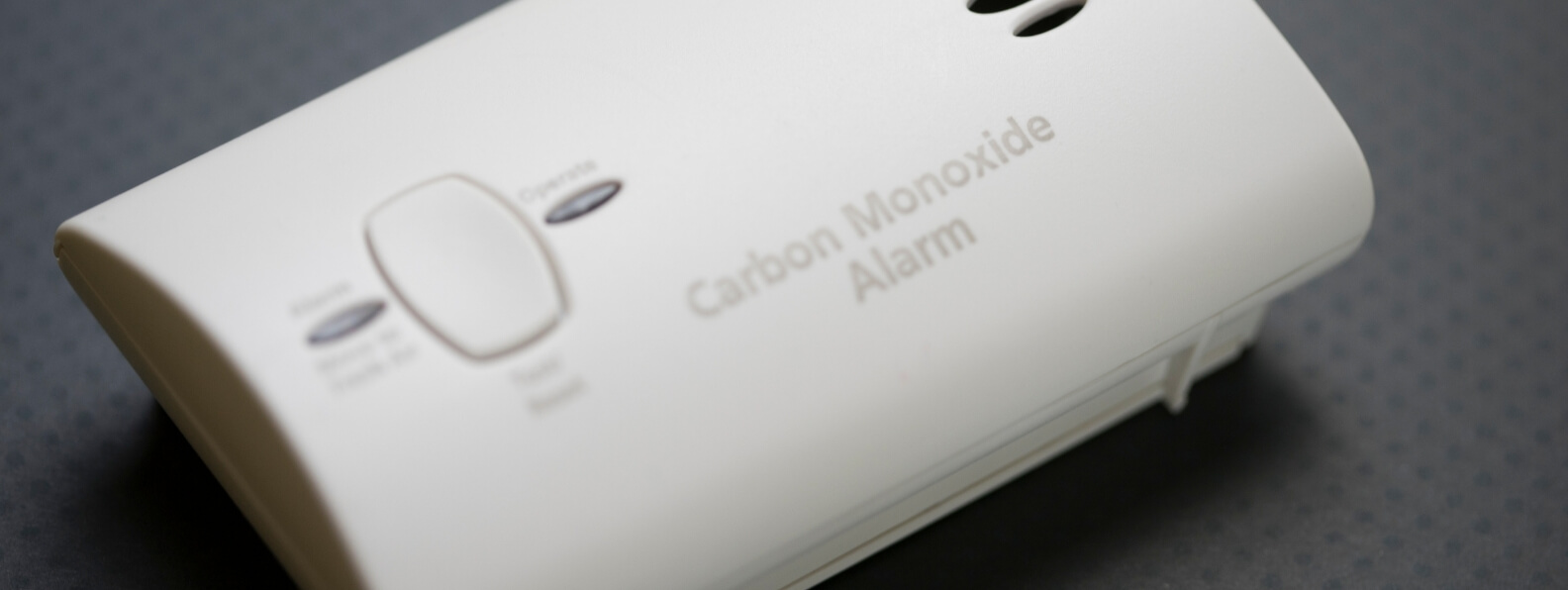
    Hyperbaric oxygen therapy, or HBOT, is the definitive treatment for clinically significant diving-related DCS. Recompression reduces bubble volume, while oxygen accelerates inert-gas elimination and supports tissues affected by impaired circulation, inflammation, and edema.

    Treatment should begin as early as practical, but a delay does not automatically eliminate the potential value of HBOT. Suspected cases require immediate oxygen, medical evaluation, consultation with a diving-medicine specialist, and coordinated transport to a suitable recompression facility. (Divers Alert Network)

    How Decompression Sickness Develops

    While a diver breathes compressed gas at depth, the increased ambient pressure causes more inert gas, usually nitrogen, to dissolve in blood and tissue. The quantity absorbed depends on depth, time, breathing-gas composition, blood flow, and tissue characteristics.

    During ascent, ambient pressure decreases and the dissolved inert gas must move from tissue into the blood, reach the lungs, and be exhaled. If pressure falls faster than the gas can be eliminated, tissues become supersaturated and bubbles may form.

    These bubbles can cause injury through several interacting mechanisms:

    • Mechanical disruption of tissue
    • Obstruction of venous or microvascular blood flow
    • Endothelial injury
    • Platelet and leukocyte activation
    • Inflammatory signaling
    • Increased vascular permeability
    • Local edema
    • Secondary tissue hypoxia

    The result is not simply a collection of gas bubbles. DCS becomes a vascular and inflammatory disorder in which tissue injury may continue even after bubble volume begins to decline. (PubMed)

    Decompression Sickness and Arterial Gas Embolism

    Decompression illness, or DCI, is an umbrella term that includes both decompression sickness and arterial gas embolism.

    DCS generally results from inert-gas bubbles forming within tissues or the venous circulation after decompression. Arterial gas embolism, or AGE, usually occurs when gas enters the arterial circulation, often following pulmonary barotrauma during ascent. AGE commonly produces abrupt stroke-like manifestations, while DCS may affect the spinal cord, brain, inner ear, joints, skin, lymphatic system, or cardiopulmonary circulation.

    The two conditions may overlap clinically, and the initial response is similar: stabilize the patient, provide the highest practical concentration of oxygen, contact emergency and diving-medicine resources, and arrange recompression when indicated. (German Journal of Sports Medicine)

    A diver who develops sudden unconsciousness, seizure, confusion, focal weakness, visual disturbance, or severe dizziness during ascent or within minutes of surfacing should be treated as having a serious decompression-related emergency. The precise distinction between DCS and AGE should not delay oxygen or evacuation.

    DCS Can Occur After an Apparently Acceptable Dive

    A history of exceeding a dive computer or decompression table strengthens suspicion, but DCS can occur after a dive that appeared to remain within accepted limits.

    Dive computers and tables estimate risk across populations. They cannot account perfectly for individual susceptibility, physiologic stress, equipment differences, or every feature of the dive.

    Factors that may increase risk include:

    • Greater depth or bottom time
    • Rapid ascent
    • Missed decompression stops
    • Repetitive or multiday diving
    • Cold exposure
    • Heavy exertion
    • Dehydration
    • Recent illness
    • Reduced physical fitness
    • Post-dive altitude exposure
    • Individual cardiovascular or pulmonary factors

    A normal dive-computer record therefore does not exclude DCS when the clinical presentation is convincing. Diagnosis depends on the dive history, timing, symptoms, examination, and exclusion of important alternative conditions. (Divers Alert Network)

    Musculoskeletal and Cutaneous Manifestations

    Musculoskeletal pain is one of the most familiar presentations. The pain may involve a shoulder, elbow, hip, knee, or another joint. It is often described as deep, poorly localized, and unrelated to movement or a specific mechanical injury.

    Skin and lymphatic findings may include:

    • Itching
    • Mottled or marbled discoloration
    • Localized swelling
    • Firm edema
    • Tender lymph nodes
    • A sense of skin crawling or abnormal sensation

    Cutis marmorata, a mottled or marbled skin pattern, should not automatically be considered a minor manifestation. It may be associated with more significant systemic involvement and warrants specialist assessment.

    Pain or skin symptoms that improve after surface oxygen may still recur. Symptom improvement should be documented, but it should not be used by itself to cancel medical evaluation or recompression planning. (PubMed)

    Neurologic Decompression Sickness

    Neurologic DCS is a serious form of decompression illness and commonly affects the spinal cord. Symptoms may begin subtly and then progress.

    Possible findings include:

    • Numbness or tingling
    • Limb weakness
    • Difficulty walking
    • Abnormal coordination
    • Loss of balance
    • Back, abdominal, or girdle-like pain
    • Altered sensation
    • Bowel or bladder dysfunction
    • Confusion
    • Changes in consciousness
    • Paralysis

    A diver may report vague fatigue, heaviness, clumsiness, or a sensation that a limb does not feel normal before an obvious deficit becomes apparent. A careful neurologic examination is therefore more useful than asking only whether the diver has pain.

    Diving-related spinal cord injury can leave persistent weakness, sensory impairment, bladder dysfunction, sexual dysfunction, and reduced mobility. Early oxygen and recompression provide the best opportunity to limit residual injury. (PubMed Central (PMC))

    Inner-Ear Decompression Sickness

    Inner-ear DCS may cause:

    • Severe vertigo
    • Nausea and vomiting
    • Imbalance
    • Nystagmus
    • Tinnitus
    • Hearing loss

    These findings can resemble inner-ear barotrauma. The distinction matters because forceful attempts to equalize pressure may worsen some barotrauma injuries, while delayed recompression may worsen DCS.

    The clinical assessment should consider the dive profile, gas mixture, timing of symptoms, difficulty equalizing during the dive, hearing changes, neurologic findings, and evidence of middle-ear injury. Consultation with a diving-medicine physician and, when available, an otolaryngologist familiar with diving injuries is appropriate.

    First responders should provide oxygen and avoid encouraging additional ear-clearing maneuvers while the diagnosis remains uncertain. (Divers Alert Network)

    Cardiopulmonary Decompression Sickness

    Severe venous bubble loads can interfere with pulmonary circulation and produce cardiopulmonary DCS, historically called “the chokes.”

    Potential findings include:

    • Cough
    • Chest pain
    • Shortness of breath
    • Rapid breathing
    • Cyanosis
    • Hypoxemia
    • Hypotension
    • Pulmonary edema
    • Cardiovascular collapse

    This is a life-threatening presentation requiring simultaneous resuscitation and urgent recompression planning. Airway management, oxygenation, hemodynamic support, and evaluation for alternative emergencies such as pneumothorax, aspiration, immersion pulmonary edema, or acute cardiac disease may take priority during initial stabilization. (PubMed Central (PMC))

    Fluid therapy should be individualized. Many divers are relatively volume depleted, but aggressive administration may worsen pulmonary edema in cardiopulmonary DCS. Management should be directed by clinicians experienced in critical care and diving medicine.

    DCS Is Primarily a Clinical Diagnosis

    There is no blood test, imaging study, or dive-computer calculation that reliably confirms or excludes DCS.

    The diagnosis is based on:

    • Exposure to reduced ambient pressure
    • The dive or pressure profile
    • The timing of symptom onset
    • The nature and distribution of symptoms
    • Neurologic and cardiopulmonary examination
    • Response to oxygen or recompression
    • Consideration of alternative diagnoses

    Laboratory testing and imaging are used primarily to evaluate severity, identify complications, and exclude other conditions.

    Potential alternatives include:

    • Stroke
    • Seizure
    • Hypoglycemia
    • Musculoskeletal injury
    • Inner-ear barotrauma
    • Pneumothorax
    • Immersion pulmonary edema
    • Contaminated breathing gas
    • Oxygen toxicity
    • Marine envenomation
    • Trauma
    • Drug or alcohol effects

    Normal imaging should not delay HBOT when a qualified clinician believes DCS is likely. Many bubble-related and microvascular injuries will not be visible on routine CT or MRI. (Divers Alert Network)

    Recording the Dive and Neurologic History

    A detailed history should be collected while emergency care continues. Useful information includes:

    • Maximum depth
    • Bottom time
    • Breathing gases
    • Ascent rate
    • Safety and decompression stops
    • Missed stops
    • Repetitive dives
    • Surface intervals
    • Previous days of diving
    • Dive-computer data
    • Exertion, cold, or equipment problems
    • Time of symptom onset
    • Symptom progression
    • Oxygen delivery and response

    A focused neurologic examination should document mental status, speech, vision, strength, sensation, coordination, gait, and balance. The examination should be repeated because findings may evolve during transport.

    Documentation should not delay evacuation. Its purpose is to preserve information that may otherwise be lost once the diver reaches an emergency department or hyperbaric center. (Divers Alert Network)

    Immediate First Aid With 100 Percent Oxygen

    The highest practical concentration of oxygen should be administered as soon as DCS is suspected.

    A demand-valve system can provide a high inspired concentration to a spontaneously breathing, cooperative patient. A nonrebreather mask may be used when a demand valve is unavailable or cannot be tolerated. A patient who is not breathing adequately requires airway support and assisted ventilation with oxygen.

    Surface oxygen may:

    • Increase oxygen delivery to injured tissue
    • Accelerate inert-gas elimination
    • Reduce bubble size by replacing nitrogen within bubbles
    • Improve symptoms before recompression
    • Limit secondary hypoxic injury

    Oxygen should generally continue during evaluation and transport, subject to available supply, airway needs, and professional direction. Symptom resolution while breathing oxygen does not prove that the injury has resolved permanently. Symptoms may recur after oxygen is discontinued. (Divers Alert Network)

    Positioning, Hydration, and Supportive Care

    The diver should be kept at rest and protected from unnecessary exertion. A comfortable supine position is generally appropriate unless vomiting, respiratory distress, trauma, or another clinical problem requires different positioning.

    The historical practice of placing suspected gas-embolism patients in a head-down position is not recommended. It can interfere with airway management and does not reliably prevent cerebral bubble distribution.

    Oral fluids may be reasonable for a fully alert patient without nausea, swallowing difficulty, or planned anesthesia. Isotonic intravenous fluids may be used when clinically appropriate, particularly if dehydration is suspected. Excessive fluid administration should be avoided in pulmonary edema, significant cardiac dysfunction, or other conditions in which fluid loading may cause harm. (German Journal of Sports Medicine)

    Routine steroids, anticoagulants, antiplatelet drugs, or other adjunctive medications should not be started solely for DCS without specialist guidance. The current U.S. Navy guidance does not recommend routine steroids or antiplatelet therapy for neurologic DCS because benefit has not been established and harm may be possible.

    Medical Stabilization Before Chamber Transfer

    A diver with severe DCS or possible AGE should generally be stabilized at the nearest capable medical facility before transport to a chamber, particularly when airway compromise, shock, trauma, pulmonary injury, or altered consciousness is present.

    This evaluation may include:

    • Airway and respiratory assessment
    • Electrocardiography
    • Glucose measurement
    • Chest imaging when pulmonary barotrauma is suspected
    • Evaluation for pneumothorax
    • Neurologic reassessment
    • Treatment of seizures or dysrhythmias
    • Management of associated trauma

    An untreated tension pneumothorax requires immediate treatment and cannot safely be ignored during recompression. Transport directly to a chamber should not replace essential resuscitation that the chamber facility cannot provide. (Divers Alert Network)

    Transport and Altitude Exposure

    Reduced atmospheric pressure at altitude can increase bubble volume and worsen decompression-related symptoms. Transport planning should therefore minimize additional altitude exposure whenever practical.

    Ground transport is often preferred when it can reach an appropriate facility without excessive delay. When air evacuation is necessary, the transport team should use the lowest practical cabin altitude or an aircraft capable of maintaining near-sea-level cabin pressure when available.

    These decisions require specialist coordination. A longer ground transfer may be less appropriate than a carefully managed flight when the patient has progressive neurologic or cardiopulmonary symptoms. Oxygen should continue during transport, and the receiving chamber should be contacted before departure. (Divers Alert Network)

    How HBOT Treats DCS

    HBOT addresses both the gas phase and the injured tissue.

    Recompression reduces bubble volume according to the relationship between pressure and gas volume. Breathing oxygen replaces nitrogen within the lungs, lowers blood nitrogen tension, and creates a gradient that favors movement of inert gas from bubbles and tissue into the circulation for elimination.

    At the same time, hyperbaric oxygen:

    • Increases dissolved plasma oxygen
    • Supports tissue beyond partially obstructed microvessels
    • Reduces edema through hyperoxic vasoconstriction
    • Improves oxygenation without proportionally reducing oxygen delivery
    • Supports cellular metabolism
    • May reduce leukocyte-endothelial interactions and inflammatory injury

    These effects explain why patients may improve rapidly during the early portion of recompression, while others continue to recover gradually after bubble reduction and tissue oxygenation have been restored. (Divers Alert Network)

    Recompression Treatment Tables

    Hyperbaric treatment is delivered according to a structured recompression profile rather than a routine outpatient wound-care protocol.

    The U.S. Navy Treatment Table 6, or a clinically equivalent oxygen recompression protocol, is widely used for diving-related DCS. Treatment commonly begins at approximately 2.8 atmospheres absolute, equivalent to 60 feet of seawater, and includes scheduled oxygen-breathing periods separated by air breaks. The profile may be extended when significant symptoms continue to improve under pressure. (UHMS)

    The selected table depends on:

    • Symptom severity
    • Neurologic findings
    • Response at treatment pressure
    • Time from symptom onset
    • Previous recompression
    • Pulmonary status
    • Oxygen tolerance
    • Chamber capability
    • The diving physician’s assessment

    More severe or refractory cases may require a longer table, a modified protocol, or additional treatments. Treatment-table selection should be directed by a physician experienced in diving and hyperbaric medicine.

    Oxygen Toxicity During Recompression

    DCS treatment tables may deliver a larger oxygen dose than many routine HBOT protocols. Scheduled air breaks help reduce uninterrupted exposure, but central nervous system or pulmonary oxygen toxicity can still occur.

    Possible warning findings include:

    • Visual or auditory changes
    • Nausea
    • Facial or limb twitching
    • Irritability
    • Dizziness
    • Seizure
    • Substernal burning
    • Cough
    • Pain with inspiration

    If central nervous system oxygen toxicity is suspected, oxygen exposure is stopped and the patient breathes chamber air according to the treatment protocol. An active convulsion should be managed without placing objects in the mouth, and decompression should wait until the patient is relaxed and breathing normally unless another overriding emergency exists.

    The possibility of oxygen toxicity does not remove the need for recompression. It requires trained chamber personnel, continuous observation, air breaks, and modification of the treatment profile when necessary.

    Early Treatment Is Preferred, but Late Treatment May Still Help

    The probability of complete recovery is generally greater when recompression begins early. Bubbles may become less responsive over time, and inflammatory or ischemic tissue injury may become established.

    Real-world treatment is often delayed because symptoms are not recognized, the diver is in a remote location, oxygen is unavailable, or transport requires many hours. A delay of 24 hours or more may reduce treatment effectiveness, but it does not prove that HBOT will provide no benefit.

    Divers with persistent or recurrent symptoms should still be discussed with a diving-medicine specialist. Meaningful improvement has been observed after delayed recompression, and the decision should be based on the patient’s current findings rather than time alone. (German Journal of Sports Medicine)

    Additional HBOT for Residual Symptoms

    Many patients improve substantially after one recompression treatment. Some have persistent weakness, sensory changes, vertigo, bladder dysfunction, pain, or cognitive findings.

    Additional HBOT may be considered when:

    • Significant symptoms remain
    • Improvement occurred during the initial treatment
    • Symptoms recur after initial relief
    • Further improvement occurs with subsequent treatment
    • Tissue remains clinically salvageable

    The U.S. Navy manual allows additional recompression for residual manifestations and advises continuing while sustained improvement is occurring. Treatment may be stopped when further sessions no longer produce meaningful progress, with the decision individualized by the treating diving-medicine physician.

    Repeat treatment should not become automatic. Persistent symptoms may also require imaging, neurologic evaluation, rehabilitation, urologic care, audiology, or assessment for another diagnosis.

    Evidence Supporting Recompression

    Recompression with oxygen is accepted as the standard treatment for DCS, although the evidence base differs from that of many common therapies.

    Randomized trials comparing recompression with no recompression are lacking. Withholding definitive treatment from a patient with neurologic, inner-ear, or cardiopulmonary DCS would create serious ethical and practical concerns. Support therefore comes from established gas physics, physiologic studies, historical experience, observational outcomes, and the frequently rapid response of symptoms during treatment. (PubMed Central (PMC))

    Randomized research has compared variations in recompression and adjunctive treatment, but there is limited evidence proving that one oxygen table is superior in every presentation. This supports individualized protocol selection while preserving the central principle that symptomatic diving-related DCS should receive hyperbaric consultation and recompression whenever feasible. (PubMed)

    In-Water Recompression Is Not Routine First Aid

    In-water recompression involves returning a symptomatic diver underwater, usually while breathing oxygen, to increase ambient pressure before controlled ascent.

    This approach carries substantial risks:

    • Drowning
    • Oxygen toxicity
    • Hypothermia
    • Vomiting or loss of consciousness underwater
    • Inadequate monitoring
    • Worsening weather or sea conditions
    • Incomplete treatment
    • Delayed evacuation

    It should not be improvised by recreational divers. In-water recompression may be considered only in exceptional remote settings where chamber evacuation is not reasonably available and where trained personnel, appropriate oxygen equipment, thermal protection, communications, and a validated protocol are already in place. (Divers Alert Network)

    Surface oxygen and organized evacuation remain the appropriate response for most diving operations.

    Observation After Recompression

    Clinical improvement at the end of a treatment does not eliminate the possibility of recurrence.

    Post-treatment observation should reflect:

    • Initial symptom severity
    • Completeness of recovery
    • Treatment table used
    • Need for repeated treatment
    • Distance from the chamber
    • Availability of a responsible companion
    • Associated medical conditions

    Current U.S. Navy guidance uses longer observation for neurologic DCS and patients requiring Treatment Table 6, and it recommends that treated patients remain near recompression capability during the early post-treatment period. Patients with residual symptoms may require hospitalization or transfer to a medical facility.

    Discharge instructions should identify recurrent pain, weakness, numbness, gait disturbance, vertigo, bladder problems, breathing difficulty, confusion, and loss of consciousness as reasons for immediate reassessment.

    Rehabilitation After Neurologic DCS

    HBOT treats the bubble-related and hypoxic components of DCS, but it cannot always reverse tissue that has already been permanently injured.

    Patients with residual neurologic impairment may require:

    • Physical therapy
    • Occupational therapy
    • Gait and balance training
    • Bladder management
    • Pain management
    • Vestibular rehabilitation
    • Audiology
    • Neuropsychological assessment
    • Psychological support

    Rehabilitation should begin as soon as the patient is medically stable. Functional recovery may continue after the final chamber treatment, particularly when the patient receives coordinated neurologic and rehabilitation care. (Divers Alert Network)

    Returning to Diving After DCS

    A diver should not return to diving solely because symptoms have disappeared.

    Assessment should consider:

    • The severity and location of the injury
    • Whether neurologic deficits completely resolved
    • The number of recompression treatments required
    • The cause of the incident
    • Pulmonary or cardiovascular risk factors
    • Possible right-to-left shunting
    • The diver’s medical fitness
    • The type of future diving planned

    A diving-medicine physician may recommend additional evaluation after neurologic, inner-ear, recurrent, or otherwise unexplained DCS. The appropriate interval before returning varies considerably and should not be determined through a universal online schedule.

    Some divers with serious residual neurologic injury, recurrent DCS, or an uncorrected predisposing condition may be advised not to return to compressed-gas diving.

    A Coordinated Clinical Response

    Effective treatment begins before the chamber doors close.

    A practical response includes:

    1. Recognize symptoms occurring after a relevant pressure exposure.
    2. Stop further diving and provide the highest practical concentration of oxygen.
    3. Perform basic stabilization and a focused neurologic assessment.
    4. Contact emergency services and a diving-medicine resource.
    5. Record the dive profile and symptom timeline.
    6. Minimize unnecessary exertion and altitude exposure.
    7. Stabilize serious airway, pulmonary, cardiac, or traumatic conditions.
    8. Arrange early recompression at a capable hyperbaric facility.
    9. Reassess after treatment and provide additional HBOT when meaningful improvement remains possible.
    10. Coordinate follow-up, rehabilitation, and future diving-clearance decisions.

    HBOT for decompression sickness is not simply oxygen treatment at a higher pressure. It is a structured emergency intervention that reduces bubble volume, accelerates inert-gas elimination, restores tissue oxygenation, and limits secondary vascular and inflammatory injury.

    Its effectiveness depends on early recognition, immediate surface oxygen, safe transport, an appropriate recompression protocol, and continued care for any residual neurologic or functional impairment.

  • Treating Carbon Monoxide Poisoning with HBOT

    Treating Carbon Monoxide Poisoning with HBOT

    Clinical Assessment, Hyperbaric Referral, Treatment Timing, Pregnancy, and Prevention of Delayed Neurologic Injury

    Carbon monoxide poisoning is a medical emergency that can injure the brain, heart, and other oxygen-dependent organs even when the patient appears to improve after leaving the exposure environment.
    Immediate treatment begins with removal from the carbon monoxide source, stabilization of the airway and circulation, and administration of 100 percent oxygen. Hyperbaric oxygen therapy, commonly abbreviated as HBOT, may then be considered for selected patients with significant neurologic, cardiac, metabolic, or exposure-related risk.

    Carbon monoxide poisoning is a recognized hyperbaric indication, but the decision to use HBOT is not based on a single laboratory value. Carboxyhemoglobin concentration, symptoms, loss of consciousness, cardiac injury, pregnancy, exposure duration, treatment delay, and the availability of a qualified hyperbaric facility must all be considered together. (CDC)

    Carbon Monoxide Causes More Than Simple Oxygen Deprivation

    Carbon monoxide is a colorless and odorless gas produced by incomplete combustion. Common exposure sources include generators, furnaces, motor vehicles, charcoal grills, propane appliances, fires, boats, and gasoline-powered tools used in enclosed or poorly ventilated areas. Multiple people from the same location may become ill at the same time. (CDC)

    Carbon monoxide binds to hemoglobin and interferes with the blood’s ability to transport oxygen. It also disrupts oxygen use and cellular respiration, particularly in organs with high metabolic requirements such as the brain and heart. This helps explain why a patient can experience serious neurologic or cardiac injury even after the measured carboxyhemoglobin level begins to decline. (CDC)

    The clinical effects may include:

    • Headache
    • Dizziness
    • Weakness
    • Nausea or vomiting
    • Chest pain
    • Shortness of breath
    • Confusion
    • Memory impairment
    • Ataxia
    • Syncope
    • Seizure
    • Coma
    • Cardiac ischemia or dysrhythmia
    • Metabolic acidosis
    • Pulmonary edema

    Symptoms are nonspecific and may resemble influenza, migraine, intoxication, gastroenteritis, stroke, or another acute medical condition. The absence of fever, a plausible combustion source, or several people with similar symptoms should raise suspicion for carbon monoxide exposure. (CDC)

    Immediate Treatment Begins Before Hyperbaric Referral

    Every patient with suspected clinically significant carbon monoxide poisoning should receive high-concentration oxygen promptly. Oxygen should not be delayed while waiting for laboratory confirmation, imaging, toxicology consultation, or acceptance by a hyperbaric facility.

    The CDC recommends administering 100 percent oxygen until the patient is symptom-free, commonly for approximately four to five hours, while performing serial neurologic examinations. Airway management, circulatory support, seizure treatment, glucose correction, trauma care, and management of cardiac or pulmonary complications should proceed simultaneously when required. (CDC)

    HBOT is an escalation of oxygen treatment rather than a replacement for emergency stabilization. A patient who requires intubation, treatment of hypotension, management of an acute coronary syndrome, or evaluation of associated trauma should receive that care before and during transfer planning.

    Early communication with a medical toxicologist, regional poison center, and hyperbaric physician can help determine whether chamber treatment is appropriate. The decision should consider both the severity of poisoning and the time required to reach a facility capable of safely managing the patient. The 2025 American College of Emergency Physicians clinical policy states that selected symptomatic patients may benefit from HBOT based on symptom severity and practical availability, including transport distance and time. (ACEP Now)

    Diagnosing Carbon Monoxide Poisoning

    Diagnosis is based on the exposure history, clinical findings, and measurement of carboxyhemoglobin through co-oximetry.

    Venous or arterial blood can be used for carboxyhemoglobin measurement. A conventional two-wavelength pulse oximeter is not reliable in carbon monoxide poisoning because it cannot accurately distinguish oxyhemoglobin from carboxyhemoglobin. A patient may therefore display a normal or reassuring pulse oxygen saturation despite significant poisoning. (CDC)

    Timing matters. Carboxyhemoglobin begins to fall after the patient leaves the exposure site and decreases more rapidly after oxygen is administered. A low measurement obtained several hours later does not exclude a clinically important exposure.

    The CDC also emphasizes that carboxyhemoglobin concentration does not correlate consistently with illness severity, clinical outcome, or response to treatment. The number should support the assessment, but it should not override neurologic findings, cardiac injury, loss of consciousness, pregnancy, or a convincing exposure history. (CDC)

    Clinical Evaluation Should Extend Beyond the COHgb Level

    A focused neurologic examination should assess mental status, memory, attention, coordination, gait, speech, motor function, and sensory findings. Brief cognitive testing can help identify abnormalities that may not be apparent during ordinary conversation. Serial examinations are useful because findings can evolve during the first several hours. (CDC)

    Additional testing may include:

    • Blood glucose
    • Electrocardiography
    • Troponin or other cardiac biomarkers
    • Blood gas and lactate assessment
    • Electrolytes and renal function
    • Toxicology testing when co-exposure is possible
    • Chest radiography in seriously poisoned patients
    • Brain CT or MRI when significant neurologic impairment, loss of consciousness, trauma, or another intracranial diagnosis is suspected

    Cardiac assessment is particularly important. Carbon monoxide can cause myocardial ischemia, ventricular dysfunction, dysrhythmia, or biomarker elevation even in patients without known coronary artery disease. The CDC notes that cardiac injury during poisoning is associated with increased long-term mortality risk, supporting ECG and cardiac biomarker evaluation in severe cases. (CDC)

    When HBOT Should Be Considered

    No single referral rule identifies every patient who will benefit. Hyperbaric consultation is commonly considered when one or more high-risk features are present.

    The CDC identifies the following considerations:

    • Carboxyhemoglobin above approximately 25 to 30 percent
    • Evidence of cardiac involvement
    • Severe metabolic acidosis
    • Transient or prolonged loss of consciousness
    • Persistent neurologic impairment
    • Abnormal neuropsychiatric testing
    • A clinical condition or exposure history suggesting severe poisoning despite a lower carboxyhemoglobin measurement
    • Pregnancy, including cases that may appear less severe in the mother

    These are consultation and treatment considerations, not absolute thresholds. A patient with a carboxyhemoglobin concentration below 25 percent may still warrant HBOT because of syncope, confusion, myocardial injury, severe acidosis, pregnancy, or prolonged exposure. Conversely, the laboratory value should be interpreted within the entire clinical picture. (CDC)

    Patient age, significant anemia, chronic cardiac disease, respiratory disease, delayed presentation, and inability to complete a reliable neurologic examination may also influence the risk assessment.

    How HBOT May Help After Carbon Monoxide Exposure

    During HBOT, the patient breathes oxygen while the chamber is pressurized above normal atmospheric pressure. This raises arterial oxygen tension and substantially increases the quantity of oxygen dissolved directly in plasma.

    The immediate treatment goals include accelerating carbon monoxide elimination, improving oxygen delivery to vulnerable tissues, and supporting cellular metabolism while hemoglobin function recovers. HBOT may also influence the inflammatory and oxidative processes believed to contribute to delayed neurologic injury after the initial hypoxic exposure has ended. (UHMS)

    The treatment is therefore intended to address more than the measured carboxyhemoglobin level. By the time the patient reaches the chamber, that level may already be much lower because of time and normobaric oxygen. The remaining clinical concern may involve ongoing brain or cardiac injury rather than the amount of carbon monoxide still circulating in the blood.

    Treatment Timing and Transfer Decisions

    When HBOT is selected, treatment is generally pursued as early as practical after stabilization. The strongest supportive randomized trial administered the hyperbaric course within 24 hours of poisoning. Delays may occur because the exposure was initially unrecognized, the patient presented late, weather limited transport, or the nearest qualified chamber was distant. (PubMed)

    Transport decisions require careful judgment. Transfer should not interrupt critical airway, cardiac, trauma, or toxicologic care. The receiving facility must be able to manage the patient’s acuity, including mechanical ventilation, vasoactive infusions, seizure risk, or associated burns when present.

    A distant transfer may provide less net benefit when the patient has mild and rapidly resolving symptoms. The balance may be different for a pregnant patient, a person with persistent neurologic impairment, or a patient with objective myocardial injury. This is why early discussion among the emergency physician, toxicologist, hyperbaric physician, transport team, and receiving center is important. (ACEP Now)

    HBOT Protocols Are Not Uniform

    Hyperbaric treatment profiles vary according to facility protocol, symptom severity, time from exposure, and the patient’s response to the first session.

    Published protocols have used initial treatment pressures between approximately 2.5 and 3.0 atmospheres absolute, frequently with scheduled air breaks. Some centers use one treatment, while others prescribe additional sessions during the first 24 hours for selected patients. The precise pressure, oxygen duration, and number of sessions should be prescribed by a hyperbaric physician rather than selected from carboxyhemoglobin concentration alone. (UHMS)

    The optimal number of sessions remains uncertain. A 2023 double-blind randomized trial compared one HBOT session with three sessions but was stopped early because of enrollment futility. It did not demonstrate a significant difference in neuropsychological sequelae between the treatment groups at six weeks or six months. These results do not compare HBOT against normobaric oxygen. They indicate that the additional benefit of two more sessions after an initial chamber treatment remains unclear. (PubMed)

    Treatment should be individualized according to the neurologic examination, cardiac status, recurrence of symptoms, treatment tolerance, and the center’s established protocol.

    Carbon Monoxide Poisoning During Pregnancy

    Pregnancy lowers the threshold for hyperbaric consultation because the fetus may be more severely affected than maternal symptoms or maternal carboxyhemoglobin levels suggest.

    The CDC recommends pregnancy testing for women of childbearing potential who are suspected of carbon monoxide poisoning. It describes HBOT as the treatment of choice for pregnant patients, including some who appear less severely poisoned, and notes that international consensus supports a more aggressive treatment approach during pregnancy. (CDC)

    Maternal stabilization remains the immediate priority because fetal oxygenation depends on maternal oxygen delivery and circulation. Obstetric consultation and fetal assessment should be incorporated when gestational age and clinical circumstances permit, but they should not delay oxygen administration.

    The decision should consider maternal symptoms, loss of consciousness, neurologic findings, fetal status, exposure duration, treatment delay, and the availability of a qualified chamber. A reassuring maternal pulse oximetry value or declining maternal carboxyhemoglobin level should not be used alone to dismiss fetal risk.

    Fire and Smoke Inhalation Require a Broader Toxicologic Assessment

    Patients exposed during a structural fire may have more than carbon monoxide poisoning. Thermal airway injury, pulmonary injury, trauma, and cyanide exposure may occur simultaneously.

    A normal chest examination early after exposure does not exclude evolving inhalation injury. The clinical team should evaluate airway burns, soot, facial injury, voice changes, respiratory distress, severe lactate elevation, altered mental status, and cardiovascular instability.

    The UHMS recognizes carbon monoxide poisoning complicated by cyanide exposure as part of the hyperbaric indication. HBOT may still be appropriate, but it does not replace airway protection, burn care, trauma resuscitation, or immediate antidotal treatment when cyanide poisoning is clinically suspected. (UHMS)

    Evidence Supporting HBOT Is Clinically Important but Mixed

    The most frequently cited supportive randomized trial was published in 2002. Symptomatic patients were assigned to three hyperbaric sessions or a comparison regimen involving normobaric oxygen and sham chamber exposures. Cognitive sequelae at six weeks occurred in approximately 25 percent of the HBOT group and 46 percent of the comparison group. The study also reported a sustained difference at 12 months. (PubMed)

    Other randomized trials have not demonstrated the same benefit. One 1999 trial found no improvement and raised the possibility of worse neuropsychological outcomes in its HBOT group, although its protocol and methodology have been the subject of substantial debate. (PubMed)

    A 2026 systematic review and meta-analysis of six randomized trials found no statistically significant overall benefit for mortality or neurologic outcomes. The authors rated the certainty of evidence as low to very low because of heterogeneity, risk of bias, differences in treatment protocols, and imprecision. (PubMed)

    The current evidence therefore does not support a claim that every patient with carbon monoxide poisoning must receive HBOT. It also does not establish that the treatment lacks value in severely poisoned or otherwise high-risk patients.

    The 2025 ACEP policy reflects this uncertainty with a Level C recommendation stating that selected symptomatic patients may benefit based on illness severity and the practical availability of treatment. (ACEP Now)

    Why the Research Remains Difficult to Interpret

    Carbon monoxide trials have used different definitions of poisoning severity, different cognitive tests, different treatment pressures, and different numbers of chamber sessions. Some enrolled mildly symptomatic patients, while others included patients with loss of consciousness or severe neurologic findings.

    Creating a credible sham is also difficult. Even small chamber pressure changes may produce physiologic effects and can reveal treatment assignment to experienced patients or staff.

    Other variables that can affect outcomes include:

    • Duration of exposure
    • Time to oxygen administration
    • Time to HBOT
    • Age
    • Cardiac injury
    • Cerebellar dysfunction
    • Co-exposure to drugs or cyanide
    • Baseline cognitive status
    • Completion of follow-up testing

    These differences help explain why systematic reviews can find substantial uncertainty even when individual trials report clinically meaningful effects.

    The evidence should be discussed honestly with patients and families. HBOT may reduce the risk of delayed cognitive injury in selected cases, but it cannot guarantee full neurologic recovery.

    Delayed Neurologic Complications

    One of the principal reasons HBOT is considered is the possibility of delayed neurologic or neuropsychiatric deterioration.

    A patient may initially recover and then develop new symptoms days or weeks later. Possible manifestations include:

    • Memory loss
    • Difficulty concentrating
    • Personality or mood change
    • Slowed thinking
    • Gait disturbance
    • Urinary incontinence
    • Parkinsonian movement
    • Weakness
    • Loss of independence

    These symptoms require medical and neurologic evaluation rather than an assumption that the patient is experiencing ordinary fatigue after hospitalization.

    HBOT delivered during the acute phase is intended partly to reduce the risk of these sequelae. The role of starting a new hyperbaric course only after delayed neurologic symptoms have already developed is less established and is supported mainly by smaller studies and case reports rather than definitive randomized evidence. (PubMed)

    Discharge Planning and Follow-Up

    Apparent symptom resolution does not eliminate the need for follow-up.

    The CDC recommends warning all discharged patients about possible delayed neurologic complications and arranging a repeat medical and neurologic examination in approximately two weeks. Earlier reassessment is appropriate when cognitive, behavioral, gait, cardiac, or respiratory symptoms develop. (CDC)

    Discharge instructions should explain that the patient should seek prompt evaluation for:

    • New confusion or memory difficulty
    • Mood or personality changes
    • Problems walking or coordinating movement
    • Recurrent headache or dizziness
    • Syncope
    • Chest pain
    • Shortness of breath
    • Weakness or seizure

    The exposure source must also be identified and corrected before anyone returns to the environment. Other people and animals from the same location may require evaluation, even when their symptoms are mild.

    Risks of HBOT

    HBOT is generally well tolerated in appropriately selected patients, but it has recognized risks.

    Potential adverse effects include:

    • Middle-ear or sinus barotrauma
    • Claustrophobia or confinement anxiety
    • Temporary visual changes
    • Pulmonary pressure injury
    • Oxygen-induced seizure
    • Rare pulmonary edema in susceptible patients

    Middle-ear barotrauma is the most common adverse effect. Oxygen-induced seizures are uncommon and generally resolve when oxygen exposure is stopped, but they require an experienced chamber team and an established emergency response protocol. (UHMS)

    An untreated pneumothorax requires correction before chamber pressurization. Additional pulmonary disease, unstable hemodynamics, implanted devices, and the requirements of mechanical ventilation should be evaluated individually.

    The safety assessment should compare these treatment risks with the risk of untreated neurologic or cardiac injury from the poisoning itself.

    Coordinating Emergency and Hyperbaric Care

    The strongest carbon monoxide treatment systems begin oxygen immediately, recognize high-risk clinical findings, and contact hyperbaric and toxicology resources early.

    A well-coordinated pathway should:

    1. Remove the patient from exposure and administer 100 percent oxygen.
    2. Stabilize the airway, breathing, and circulation.
    3. Confirm exposure with co-oximetry without relying on conventional pulse oximetry.
    4. Perform serial neurologic and cardiac assessment.
    5. Identify pregnancy, loss of consciousness, severe acidosis, neurologic impairment, or myocardial injury.
    6. Consult a poison center, toxicologist, and hyperbaric physician when high-risk features are present.
    7. Balance expected benefit against transport delay and the patient’s stability.
    8. Provide follow-up for delayed neurologic and cardiac complications.

    HBOT should be presented neither as mandatory for every exposure nor as unnecessary because randomized evidence is inconsistent. It is a time-sensitive treatment option for selected patients whose neurologic, cardiac, maternal, fetal, metabolic, or exposure-related risk justifies the additional intervention.

    The decision is strongest when it is made through clinical assessment rather than a laboratory threshold alone.

  • Current Research in Hyperbaric Medicine

    Current Research in Hyperbaric Medicine

    Where the Evidence Is Advancing, Which Questions Remain Unanswered, and How Modern Studies Are Refining HBOT

    Hyperbaric oxygen therapy, commonly abbreviated as HBOT, has established roles in conditions such as decompression sickness, arterial gas embolism, carbon monoxide poisoning, delayed radiation injury, selected advanced diabetic foot ulcers, refractory osteomyelitis, and compromised grafts or flaps.

    Current research is moving beyond the broad question of whether oxygen under pressure produces biologic effects. Those effects are already well documented. Investigators are increasingly asking more clinically useful questions:

    • Which patients are most likely to benefit?
    • How early should treatment begin?
    • What pressure and oxygen duration provide the best balance of benefit and risk?
    • Which outcomes should determine whether treatment continues?
    • Can biomarkers or imaging predict response?
    • Are improvements durable after the treatment course ends?

    The answers are indication specific. Research supporting HBOT for radiation cystitis cannot automatically be applied to traumatic brain injury, diabetic wounds, long COVID, or routine postoperative recovery. Each condition requires its own treatment rationale, comparison group, dosing strategy, and patient-centered outcomes.

    Late Radiation Injury Is Producing Some of the Strongest New Evidence

    Delayed radiation injury remains one of the most active areas of hyperbaric research. Radiation can progressively damage small blood vessels, reduce capillary density, increase fibrosis, and leave previously treated tissues chronically hypoxic.

    The RICH-ART randomized trial previously demonstrated that HBOT improved patient-reported urinary symptoms in patients with chronic radiation cystitis. Five-year follow-up published in 2025 found that symptom improvements remained clinically meaningful among many initial responders. Of the 70 patients included in the long-term analysis, 48 met the study’s responder criteria after treatment. Some patients received an additional course when symptoms recurred. (PubMed)

    These long-term findings are important because HBOT is intended to stimulate tissue and vascular remodeling, not simply produce temporary symptom relief during chamber exposure. The study also identified several priorities for future research, including optimal treatment protocols, predictive biomarkers, and health-economic evaluation. (PubMed)

    Research involving late radiation effects after breast cancer treatment has produced more nuanced results. In a 2024 randomized clinical trial, offering HBOT did not significantly reduce pain in the intention-to-treat analysis, but it did reduce fibrosis. Among the smaller group of participants who completed treatment, both pain and fibrosis improved. Low treatment uptake demonstrated that a therapy can appear biologically effective while remaining difficult for patients to complete because of time, travel, or treatment burden. (PubMed)

    A 2025 systematic review of comparative studies involving late bowel and bladder radiation injury continued to find evidence of benefit in selected patients while emphasizing heterogeneity in patient selection, protocols, and outcome measurement. (PubMed)

    The research direction is therefore shifting from treating all delayed radiation complications as one category toward identifying which organs, symptoms, injury patterns, and stages respond most reliably.

    Researchers Are Examining the Optimal Hyperbaric Oxygen Dose

    A hyperbaric treatment is defined by more than the presence of oxygen. Pressure, oxygen-breathing time, air breaks, session frequency, and total treatment number all contribute to the dose.

    Many established protocols were developed through clinical experience, physiologic reasoning, and institutional practice rather than direct randomized comparisons of different pressure levels. Current research is beginning to evaluate whether lower or higher pressures produce meaningful differences in outcomes and adverse effects.

    A 2026 retrospective comparison of HBOT at 2.0 and 2.5 atmospheres absolute for hemorrhagic radiation cystitis found that fewer patients treated at 2.5 ATA experienced gross hematuria within one year. However, the time to recurrence and other urinary symptoms were similar, while adverse events were more frequent at the higher pressure. Because the study was retrospective and involved treatment at two different sites, randomized research is still needed before concluding that one pressure is superior. (Wiley Online Library)

    This type of research reflects a central principle of modern hyperbaric medicine: more pressure is not automatically more therapeutic.

    A higher oxygen partial pressure may increase tissue oxygenation and selected cellular responses, but it may also increase barotrauma, oxygen-toxicity risk, visual effects, and treatment intolerance. A lower-pressure protocol may be easier to tolerate but could require longer oxygen periods or more sessions to achieve a comparable biologic exposure.

    Future comparative trials will need to evaluate several outcomes simultaneously:

    • Symptom response
    • Durability of benefit
    • Adverse effects
    • Treatment completion
    • Number of sessions required
    • Cost and resource utilization
    • Patient quality of life

    The most effective protocol may differ by diagnosis. The oxygen dose needed for an acute gas embolism is not necessarily the appropriate dose for a chronic radiation injury or diabetic foot ulcer.

    Patient Selection Remains Central to Diabetic Wound Research

    Research involving diabetic foot ulcers continues to produce mixed results because the term diabetic foot ulcer includes wounds with very different causes and healing potential.

    Some ulcers are primarily neuropathic and exposed to ongoing pressure. Others are ischemic, infected, deeply invasive, or complicated by osteomyelitis. Studies also differ in vascular assessment, offloading quality, wound severity, surgical care, and definitions of healing or amputation.

    Current research increasingly recognizes that HBOT should not be studied as a replacement for comprehensive diabetic foot care. It must be evaluated alongside:

    • Effective pressure offloading
    • Vascular assessment and revascularization
    • Debridement
    • Infection treatment
    • Glucose management
    • Nutritional support
    • Appropriate wound dressings

    The most clinically relevant question is not whether HBOT helps an average diabetic wound. It is whether it improves healing or limb preservation in a defined subgroup whose tissue remains hypoxic despite appropriately delivered standard care.

    Modern studies are also exploring methods of identifying that subgroup. Transcutaneous oxygen measurement, toe pressure, Doppler waveforms, wound classification, perfusion imaging, and the wound’s early response to standard treatment may all contribute to more precise patient selection.

    This precision is essential because a treatment can appear ineffective when applied to patients whose primary barrier is not oxygen responsive. HBOT cannot compensate for untreated arterial obstruction, continued pressure, retained necrotic tissue, or an undrained infection.

    Infection Research Is Focused on Timing and Treatment Integration

    Necrotizing soft tissue infections remain an important but difficult area of HBOT research.

    The physiologic rationale includes increased oxygen tension in hypoxic tissue, support of leukocyte microbial killing, inhibition of selected anaerobic organisms, reduction of edema, and modulation of inflammatory injury. However, these infections require immediate surgery, antimicrobial treatment, and critical care. Randomized trials are difficult because the condition is uncommon, rapidly progressive, and clinically heterogeneous.

    A 2025 prospective Scandinavian observational study found an association between HBOT and lower 30-day mortality. However, patients who received HBOT were less acutely ill at baseline, creating a substantial risk of selection bias. The investigators concluded that randomized research is still needed to determine whether HBOT itself caused the observed outcome difference. (PubMed)

    Another 2025 study reported a stronger apparent mortality association among patients with larger wounds and higher APACHE II severity scores. These findings may help generate hypotheses about which patients should be prioritized in future trials, but they do not establish definitive treatment criteria. (PubMed)

    Current infection research is therefore addressing several operational questions:

    • How soon after the first debridement should HBOT begin?
    • Which microbial or clinical patterns are most oxygen responsive?
    • Should treatment be delivered once or more than once daily during the acute phase?
    • Can HBOT reduce the amount of tissue removed during later operations?
    • Does treatment improve survival independently of specialized-center care?

    Until these questions are resolved, HBOT remains an adjunct. It must never delay surgical source control, antibiotic administration, resuscitation, or a necessary return to the operating room.

    Trauma Research Is Moving Toward Function, Not Only Limb Survival

    Earlier trauma research often focused on amputation, infection, or visible tissue necrosis. Newer studies increasingly recognize that long-term function may be equally important.

    Severe crush injuries and open fractures can leave a limb technically intact but affected by chronic pain, stiffness, weakness, nerve injury, infection, repeated surgery, or poor quality of life. Research must therefore measure more than whether amputation occurred.

    Important outcomes include:

    • Amount of tissue necrosis
    • Number of repeat operations
    • Infection and osteomyelitis
    • Fracture healing
    • Need for reconstruction
    • Pain and mobility
    • Return to work
    • Long-term limb function

    This broader approach may identify meaningful benefits that a single short-term composite endpoint does not capture. It also prevents researchers from overstating success when a limb is preserved but remains severely impaired.

    Future trials in acute traumatic ischemia will need to coordinate HBOT tightly with vascular repair, fasciotomy, fixation, debridement, and reconstructive surgery. Timing is likely to matter because HBOT is most biologically plausible while threatened tissue remains viable.

    Neurologic Research Remains Promising but Investigational

    The brain is a major focus of emerging hyperbaric research. Proposed mechanisms include improved oxygen availability, altered cerebral blood flow, angiogenic signaling, neuroplasticity, mitochondrial effects, and modulation of neuroinflammation.

    These mechanisms have led investigators to study HBOT in persistent traumatic brain injury symptoms, post-concussion syndromes, stroke recovery, cognitive decline, and other neurologic conditions.

    The evidence remains unsettled. Neurologic trials are especially difficult to interpret because pressurized air may not function as a completely inactive placebo. Even modest increases in pressure can alter gas partial pressures and produce physiologic effects. Symptoms such as fatigue, concentration difficulty, headache, sleep disturbance, and mood change are also influenced by rehabilitation, expectation, time, and concurrent treatment.

    A registered blinded, adaptive, randomized, placebo-controlled trial is currently evaluating HBOT for mild to moderate traumatic brain injury in veterans and active-duty military personnel. The study is designed to address persistent uncertainty using stronger masking and adaptive trial methods. (ClinicalTrials.gov)

    Until sufficiently rigorous trials demonstrate reproducible clinical benefit, neurologic uses outside recognized indications should be described as investigational. Mechanistic plausibility and changes on functional imaging do not automatically establish improved daily function.

    Long COVID Research Illustrates Why Controlled Trials Matter

    Early studies of HBOT for post-COVID conditions reported improvements in fatigue, cognition, sleep, pain, and quality of life. These findings generated interest because long COVID may involve endothelial dysfunction, inflammation, altered oxygen utilization, autonomic disturbance, and neurologic symptoms. (PubMed)

    A 2025 randomized, double-blind, placebo-controlled phase 2 trial produced a more cautious result. Eighty participants received either 10 HBOT sessions or placebo treatment. Both groups improved, but there was no significant difference between HBOT and placebo in the primary short-term physical-function outcomes. (PubMed)

    This trial does not prove that every possible hyperbaric protocol is ineffective for long COVID. It evaluated a defined course of 10 treatments in a particular population. However, it shows why uncontrolled improvement cannot automatically be attributed to HBOT.

    Researchers still need to determine whether:

    • A specific long COVID phenotype is more responsive
    • More than 10 sessions are required
    • Benefits are cognitive, autonomic, respiratory, or systemic
    • Improvements persist after treatment
    • The treatment is cost effective
    • A truly credible sham protocol can be designed

    Feasibility studies and additional registered trials are continuing to examine whether larger, well-controlled research is practical. (ClinicalTrials.gov)

    For now, HBOT for long COVID should remain within appropriately reviewed research or carefully defined investigational settings rather than being represented as established therapy.

    Biomarkers May Help Predict Who Will Respond

    One of the most important research directions is the development of predictive biomarkers.

    Current hyperbaric practice often relies on diagnosis, wound classification, clinical examination, imaging, tissue oxygen testing, and response over time. These methods are useful but do not always predict which individual patient will complete treatment successfully.

    Potential research targets include:

    • Transcutaneous oxygen response
    • Endothelial and angiogenic markers
    • Inflammatory cytokines
    • Circulating progenitor cells
    • Tissue perfusion imaging
    • Microvascular density
    • Metabolic or mitochondrial markers
    • Baseline symptom severity
    • Time from injury to treatment

    A useful biomarker must do more than change after HBOT. It should predict a clinically meaningful outcome, such as wound closure, cessation of bleeding, tissue salvage, improved function, or reduced need for surgery.

    Researchers must also distinguish between markers of oxygen exposure and markers of therapeutic response. An increase in arterial or tissue oxygen confirms that the dose reached the patient. It does not prove that the underlying disease process improved.

    Patient-Reported Outcomes Are Becoming More Important

    Traditional hyperbaric research frequently measured wound size, amputation, survival, or visible bleeding. These outcomes remain important, but they do not fully describe how treatment affects a patient’s life.

    A patient with radiation cystitis may value uninterrupted sleep, freedom from clot retention, or the ability to travel without fear of bleeding. A person with late breast radiation injury may value reduced tissue tightness and improved shoulder movement. A trauma survivor may prioritize walking ability and return to work over the appearance of the wound.

    Current and future studies increasingly need to include:

    • Pain
    • Mobility
    • Urinary or bowel function
    • Fatigue
    • Sleep
    • Cognitive function
    • Independence
    • Return to work
    • Treatment burden
    • Quality of life

    Patient-reported outcomes were central to the RICH-ART research and helped demonstrate that HBOT’s effects on chronic radiation cystitis extended beyond a simple yes-or-no assessment of hematuria. (PubMed)

    Research Must Account for Treatment Burden and Access

    HBOT often requires attendance five days per week for several weeks. A scientifically effective treatment may still have limited real-world value when patients cannot access or complete it.

    Research should therefore evaluate:

    • Travel distance
    • Missed work
    • Caregiver requirements
    • Transportation
    • Insurance authorization
    • Treatment discontinuation
    • Chamber availability
    • Cost per meaningful clinical outcome

    The 2024 breast radiation trial demonstrated this challenge clearly. Only a minority of patients offered HBOT completed treatment, complicating interpretation of both effectiveness and real-world feasibility. (PubMed)

    Health-economic research is particularly important for long treatment courses. Investigators must determine whether the cost of HBOT is offset by reductions in hospitalization, transfusion, surgery, amputation, long-term wound care, or loss of function.

    The Next Phase of Hyperbaric Research

    The field does not need more studies that group unrelated diagnoses together or measure only whether a biologic marker changed after oxygen exposure.

    The highest-priority research questions are increasingly clear:

    • Which patient phenotypes respond to HBOT?
    • What is the minimum effective oxygen dose?
    • When should treatment begin?
    • Which standard therapies must be completed first?
    • What outcomes justify continuing treatment?
    • How durable are the benefits?
    • Which adverse effects are pressure dependent?
    • Can biomarkers guide treatment selection?
    • Is HBOT cost effective compared with available alternatives?

    Current research supports a disciplined view of hyperbaric medicine. Evidence continues to strengthen for selected delayed radiation injuries, while dose optimization and long-term outcomes remain active areas of study. Observational findings in severe infection are encouraging but require randomized confirmation. Neurologic conditions and long COVID remain investigational, with controlled trials producing mixed or negative results.

    This is how a medical specialty matures. The objective is not to prove that HBOT works for everything or to dismiss it because evidence varies among indications. The objective is to identify where oxygen under pressure produces measurable, durable, and patient-important benefits, then deliver it at the safest and most effective dose.

  • Oxygen Toxicity in Hyperbaric Medicine

    Oxygen Toxicity in Hyperbaric Medicine

    Understanding Central Nervous System Effects, Pulmonary Exposure, Risk Factors, Prevention, and Emergency Response

    Oxygen is essential to human metabolism, but it also behaves like a medication. Its physiologic effects depend on the concentration delivered, the surrounding pressure, the duration of exposure, and the individual patient’s susceptibility.

    During hyperbaric oxygen therapy, or HBOT, a patient breathes oxygen at an ambient pressure greater than normal atmospheric pressure. This produces oxygen partial pressures that cannot be reached by breathing supplemental oxygen at sea level alone.

    The resulting hyperoxia is responsible for many of HBOT’s therapeutic effects. It increases dissolved plasma oxygen, supports oxygen-dependent immune activity, influences vascular signaling, and improves diffusion into selected hypoxic tissues. The same exposure can become toxic when the oxygen dose exceeds the capacity of cellular antioxidant systems.

    Modern clinical protocols are designed to remain within established therapeutic limits. Serious oxygen toxicity is uncommon during medically supervised HBOT, but every hyperbaric team must understand how to recognize, prevent, and manage it.

    Oxygen Toxicity Is a Dose-Dependent Effect

    Oxygen toxicity is not an allergy to oxygen. It is a dose-related cellular response to elevated oxygen partial pressure.

    The oxygen dose delivered during HBOT is influenced by:

    • Treatment pressure
    • Oxygen concentration
    • Duration of each oxygen-breathing period
    • Length and frequency of air breaks
    • Number of treatments
    • Interval between treatments
    • Patient-specific physiologic factors

    A higher pressure increases the partial pressure of oxygen delivered to the lungs. Longer uninterrupted exposure increases the time during which tissues are exposed to that pressure. Repeated sessions increase cumulative exposure, particularly in the lungs and ocular tissues.

    The two principal clinical patterns are:

    • Central nervous system oxygen toxicity, which is generally associated with shorter exposure to high oxygen partial pressures and may culminate in a seizure
    • Pulmonary oxygen toxicity, which is more closely associated with prolonged or cumulative oxygen exposure and may cause respiratory irritation or reduced lung function

    These patterns overlap, but they occur on different exposure timelines. Acute central nervous system effects can develop within a single treatment, while clinically significant pulmonary toxicity is more often associated with longer or unusually intensive oxygen schedules. (PubMed)

    Reactive Oxygen Species and Cellular Injury

    Normal oxygen metabolism produces reactive oxygen species, including superoxide and hydrogen peroxide. These molecules are not inherently harmful. At controlled levels, they participate in cell signaling, immune function, vascular regulation, and adaptation to physiologic stress.

    Cells protect themselves through antioxidant systems such as superoxide dismutase, catalase, glutathione, and other enzymatic and nonenzymatic defenses.

    Hyperbaric exposure increases the generation of reactive oxygen and nitrogen species. When production temporarily exceeds the ability of antioxidant systems to neutralize them, oxidative stress can affect cell membranes, proteins, enzymes, mitochondria, and signaling pathways.

    In the central nervous system, this imbalance may alter neuronal excitability and inhibitory signaling. In the lungs, repeated oxidative injury may affect the alveolar-capillary membrane and pulmonary epithelium. The precise sequence leading from therapeutic hyperoxia to clinical toxicity remains incompletely understood and varies among patients. (PubMed)

    This biologic variability explains why oxygen toxicity cannot be predicted from pressure and exposure time alone. Two patients receiving the same protocol may not have the same response, and an individual patient’s tolerance may vary from one day to another.

    Central Nervous System Oxygen Toxicity

    Central nervous system oxygen toxicity, often abbreviated as CNS oxygen toxicity, is the most dramatic acute oxygen-related complication of HBOT.

    Its most recognizable manifestation is a generalized tonic-clonic seizure. These events are uncommon during standard clinical protocols and are usually self-limited after the patient is removed from oxygen exposure. They still require immediate, coordinated management because the chamber environment limits access and introduces additional airway and pressure-related concerns.

    Possible warning symptoms include:

    • Visual changes, such as narrowing of the visual field or unusual lights
    • Ringing in the ears or other auditory changes
    • Nausea
    • Facial, lip, or limb twitching
    • Irritability, agitation, confusion, or unusual behavior
    • Dizziness
    • Tingling sensations
    • A sense of apprehension
    • Generalized seizure

    These symptoms are sometimes organized into the mnemonic VENTID-C: visual changes, ear symptoms, nausea, twitching, irritability, dizziness, and convulsion.

    The mnemonic is useful for staff education, but it should not create false reassurance. Warning symptoms are inconsistent, and a seizure may occur without a recognizable prodrome. (NCBI)

    Not every seizure occurring inside a hyperbaric chamber is caused by oxygen toxicity. Hypoglycemia, fever, medication effects, withdrawal, intracranial disease, carbon monoxide poisoning, and a preexisting seizure disorder may produce similar events. Published case reports describe seizure-like episodes during HBOT that were ultimately attributed to causes other than CNS oxygen toxicity. (PubMed)

    A post-event evaluation should therefore investigate alternative or contributing causes rather than automatically labeling every chamber seizure as an oxygen reaction.

    How Common Are Oxygen Toxicity Seizures?

    Reported seizure rates vary according to treatment pressure, oxygen duration, air-break practices, patient population, indication, and how events are classified.

    Large clinical series consistently indicate that seizures are uncommon. One review of more than 80,000 treatments reported two seizures, or approximately 2.4 events per 100,000 treatments. Another analysis of 62,614 sessions identified seven seizures from all causes, with only one considered clearly attributable to oxygen toxicity. (PubMed)

    A larger study examining more than 180,000 treatments found a seizure frequency of 3.9 per 10,000 treatments before the introduction of a five-minute air break and 1.2 per 10,000 treatments afterward. Differences between studies illustrate how protocol design and event classification affect reported incidence. (PubMed)

    Patients should be informed that the risk exists, but it should be presented in proportion. For most appropriately screened patients receiving standard clinical protocols, a CNS oxygen toxicity seizure is a rare event.

    Factors That May Lower the Seizure Threshold

    The strongest treatment-related risk factors are increased oxygen partial pressure and longer uninterrupted oxygen exposure. Other physiologic or clinical conditions may reduce an individual patient’s tolerance.

    Potential contributors include:

    • Fever
    • Hypoglycemia
    • Hypercapnia or carbon dioxide retention
    • Significant respiratory obstruction
    • Uncontrolled seizure disorder
    • Acute brain injury
    • Alcohol or sedative withdrawal
    • Stimulant or recreational drug exposure
    • Certain medications that influence seizure threshold
    • Severe physiologic stress
    • Prolonged or repeated high-pressure oxygen exposure

    Carbon dioxide is particularly relevant because it increases cerebral blood flow and may increase oxygen delivery to the brain. A patient who is hypoventilating, retaining carbon dioxide, exercising under pressure, or breathing through excessive apparatus resistance may theoretically reach a toxic cerebral oxygen exposure more quickly.

    The Undersea and Hyperbaric Medical Society notes that fever, low blood glucose, medications, and underlying disease processes may lower the threshold for oxygen toxicity in some patients. These factors should be assessed clinically, but they are not precise predictors of who will experience a seizure. (UHMS)

    A history of seizures is generally treated as a relative consideration rather than an automatic exclusion. The hyperbaric physician should review seizure control, medication adherence, recent events, metabolic abnormalities, fever, and the urgency of the HBOT indication.

    Responding to a Seizure Under Pressure

    A seizure during HBOT requires an immediate but controlled response.

    The first action is to discontinue oxygen exposure. In a multiplace chamber, the patient’s oxygen mask or hood is removed so the patient breathes the chamber’s compressed-air atmosphere. In a monoplace chamber, air may be supplied through the designated breathing system when the chamber design and protocol permit.

    The treatment team should then:

    • Protect the patient from striking the chamber or equipment
    • Maintain the airway as safely as possible
    • Avoid placing objects in the patient’s mouth
    • Observe breathing and circulation
    • Prepare for suction, airway support, or emergency medication if needed
    • Check glucose and assess other possible causes after the event
    • Follow the facility’s emergency protocol and physician direction

    The chamber should not be rapidly decompressed during an active generalized convulsion. A convulsing patient may briefly hold their breath or have an obstructed airway. Expanding pulmonary gas during decompression could create an additional risk of pulmonary barotrauma. Hyperbaric nursing guidance advises waiting for the convulsion to stop and spontaneous respiration to return before beginning controlled decompression. (UHMS)

    Once oxygen exposure is stopped, an oxygen toxicity seizure commonly resolves without anticonvulsant medication. Medication may still be required when seizure activity is prolonged, recurrent, or suspected to have another cause.

    The patient should receive a full medical assessment before any decision is made about resuming HBOT. A treatment may later continue at a reduced pressure, with shorter oxygen periods, additional air breaks, or after correction of a contributing factor. The decision depends on the indication, event severity, and expected benefit of continued therapy.

    Pulmonary Oxygen Toxicity

    The lungs are continuously exposed to the inspired treatment gas, making them an important site of cumulative oxygen effects.

    Pulmonary oxygen toxicity begins with oxidative irritation of the respiratory epithelium and alveolar-capillary interface. With sufficient exposure, patients may develop:

    • Dry cough
    • Substernal burning or discomfort
    • Chest tightness
    • Pain with deep inspiration
    • Shortness of breath
    • Reduced exercise tolerance
    • A measurable decline in vital capacity

    More advanced pulmonary injury is uncommon in routine outpatient HBOT. It is more relevant during prolonged treatment tables, repeated emergency treatments, intensive diving exposures, critical-care oxygen therapy, or other situations involving a high cumulative oxygen dose.

    A 2023 longitudinal study evaluated patients receiving 100 percent oxygen at 2.0 to 2.4 ATA for 90 minutes, five days per week. Investigators found no significant changes in measured pulmonary function after 20, 40, or 60 treatments, including among patients with preexisting respiratory disease. These findings support the pulmonary safety of commonly used modern protocols while not eliminating the need to evaluate new respiratory symptoms. (PubMed)

    Patients who develop persistent cough, chest discomfort, new dyspnea, or an unexplained change in respiratory function should be reassessed. The treatment pressure, exposure time, cumulative dose, pulmonary history, and alternative diagnoses should be reviewed.

    Pulmonary Disease and Oxygen Toxicity Are Not the Same Risk

    Pulmonary oxygen toxicity should be distinguished from pulmonary barotrauma.

    Oxygen toxicity is caused by the biochemical effects of elevated oxygen exposure. Pulmonary barotrauma is caused by the expansion or compression of gas within the lungs during pressure changes.

    A patient with airway obstruction, bullous disease, pulmonary air trapping, or a history of spontaneous pneumothorax may be at increased risk for pressure-related injury during decompression. This does not necessarily mean the patient has an increased biochemical susceptibility to pulmonary oxygen toxicity.

    The risks can overlap in the same patient, but they require different screening and preventive strategies. UHMS guidance identifies significant air trapping, airway obstruction, and previous spontaneous pneumothorax as concerns requiring a careful benefit-risk assessment before HBOT. (UHMS)

    An untreated pneumothorax remains a major contraindication because trapped pleural gas may expand during decompression.

    Ocular Effects of Repeated Oxygen Exposure

    Repeated HBOT may produce temporary changes in the eye, most commonly a myopic shift. Patients may notice blurred distance vision while near vision remains stable or improves.

    This change is associated with oxygen-related alterations in the crystalline lens rather than injury to the retina or optic nerve in routine treatment. It often develops gradually during a multweek course and usually improves after treatments are completed.

    Very prolonged cumulative exposure has also been associated with cataract development or progression. These effects are distinct from acute CNS oxygen toxicity but remain part of the broader discussion of cumulative oxygen dose. (Mayo Clinic)

    Patients should report sudden visual loss, eye pain, flashes, floaters, or major visual-field changes promptly. These findings should not be assumed to represent the expected temporary refractive shift.

    Air Breaks and Oxygen Toxicity Prevention

    An air break is a planned interval during which the patient stops breathing oxygen but remains at treatment pressure.

    In a multiplace chamber, the patient generally removes the oxygen mask or hood and breathes the compressed-air chamber atmosphere. In a monoplace chamber, air is delivered through an approved breathing system.

    Air breaks are used to interrupt continuous oxygen exposure and reduce the likelihood of central nervous system and pulmonary oxygen toxicity. They are commonly included in protocols delivered above 2 ATA or when treatment duration and patient risk justify them. (UHMS)

    The large retrospective study involving more than 180,000 sessions found that introduction of a five-minute air break was associated with a significant reduction in seizure frequency. Because the study was observational, it cannot prove that the air break alone caused the reduction, but it supports their practical role in clinical protocols. (PubMed)

    An air break does not eliminate all risk. A seizure can still occur after an air break or during a protocol that has been tolerated previously. The complete oxygen dose and the patient’s current clinical condition remain important.

    Treatment Protocols Are Designed Around Oxygen Dose

    Hyperbaric protocols intentionally control pressure and exposure duration. A standard treatment may divide the oxygen dose into several breathing periods separated by air breaks rather than providing one uninterrupted exposure.

    Risk-reduction strategies may include:

    • Selecting the lowest effective treatment pressure for the indication
    • Limiting uninterrupted oxygen-breathing periods
    • Incorporating scheduled air breaks
    • Avoiding unnecessary extension of treatment time
    • Reassessing unusually intensive or twice-daily schedules
    • Reviewing medications and recent clinical changes
    • Correcting fever or hypoglycemia before treatment
    • Monitoring patients continuously
    • Adjusting future treatments after suspected toxicity

    More oxygen does not automatically produce a better clinical outcome. Increasing pressure or duration may increase toxicity risk without increasing therapeutic benefit.

    Protocols should be selected for the diagnosis rather than copied from another indication. A treatment table used for decompression illness or arterial gas embolism may involve a different exposure pattern from a routine wound or radiation-injury protocol.

    Pre-Treatment Screening Reduces Avoidable Risk

    Screening should occur before the treatment course and continue before every session.

    The clinical team should review:

    • New fever or infection symptoms
    • Recent seizure activity
    • Medication additions or interruptions
    • Alcohol, sedative, or stimulant withdrawal
    • Blood glucose when clinically indicated
    • Changes in respiratory status
    • New confusion, weakness, or neurologic symptoms
    • Sleep deprivation or severe physiologic stress
    • Previous difficulty tolerating oxygen exposure
    • Recent treatment interruptions or adverse events

    A stable patient can develop a new risk factor during a long treatment course. A respiratory infection, medication change, poor oral intake, or loss of glucose control may alter the safety of that day’s treatment.

    Patients should be encouraged to report symptoms honestly. They should not feel that mentioning nausea, twitching, visual changes, or anxiety will be interpreted as failure to cooperate.

    Continuous Observation Is Essential

    Oxygen toxicity cannot be prevented through screening alone. Staff must observe the patient throughout compression, oxygen breathing, air breaks, and decompression.

    Observation should include:

    • Facial movement and twitching
    • Behavior and mental status
    • Communication ability
    • Respiratory pattern
    • Reports of nausea, dizziness, or visual change
    • Oxygen-delivery system performance
    • Glucose-related symptoms
    • Changes in monitoring data when physiologic monitoring is used

    In a monoplace chamber, observation and communication occur through the transparent chamber wall and intercommunication system. In a multiplace chamber, an inside attendant may provide direct assessment while communicating with the chamber operator and attending clinician.

    The FDA advises facilities to monitor and supervise patients throughout treatment, maintain trained staff, follow manufacturer instructions, and perform the required equipment maintenance and safety checks. (U.S. Food and Drug Administration)

    Patient Counseling Should Be Accurate and Reassuring

    Patients deserve a clear explanation of oxygen toxicity without language that creates unnecessary fear.

    They should understand that:

    • Oxygen is being delivered at a controlled medical dose.
    • Serious oxygen toxicity is uncommon during standard clinical HBOT.
    • Staff monitor continuously for early symptoms.
    • Air breaks may be used to reduce uninterrupted oxygen exposure.
    • A seizure is possible but rare.
    • New cough, chest discomfort, visual changes, twitching, nausea, or unusual sensations should be reported immediately.
    • Treatment can be stopped or modified when a safety concern develops.

    Patients should also be told that a previous uneventful session does not guarantee that every later treatment will feel identical. Daily communication remains important throughout the course.

    Oxygen toxicity represents the point at which the therapeutic oxygen dose exceeds an individual tissue’s tolerance. In clinical hyperbaric medicine, that risk is managed through appropriate pressure selection, limited exposure periods, air breaks, patient screening, continuous observation, and rehearsed emergency procedures.

    The objective is not to avoid oxygen’s biologic effects. Those effects are the reason HBOT is prescribed. The objective is to deliver enough oxygen to achieve the intended clinical response while remaining within a carefully controlled safety range.

  • HBOT and Angiogenesis

    HBOT and Angiogenesis

    How Hyperbaric Oxygen Therapy Influences New Blood Vessel Formation, Cellular Signaling, and Tissue Repair

    Angiogenesis is the formation of new capillary branches from existing blood vessels. It is a critical part of wound healing because newly repaired tissue requires a dependable microvascular network to deliver oxygen, nutrients, immune cells, and signaling molecules.

    Hyperbaric oxygen therapy, commonly abbreviated as HBOT, may support this process by temporarily increasing tissue oxygen tension and influencing several pathways involved in vascular repair. These include vascular endothelial growth factor, nitric oxide signaling, endothelial cell activity, and the mobilization of circulating progenitor cells.

    The relationship is more complex than the simple statement that oxygen grows blood vessels. Angiogenesis depends on coordinated cycles of signaling, vessel sprouting, extracellular matrix remodeling, blood flow, and vessel maturation. HBOT may influence several of these steps, but it cannot restore circulation through a completely obstructed artery or create viable tissue where irreversible necrosis has already occurred.

    Angiogenesis and Vasculogenesis Are Related but Distinct

    Angiogenesis describes the growth of new capillary branches from blood vessels already present within the tissue. Endothelial cells lining an existing vessel become activated, migrate into the surrounding matrix, proliferate, and organize into new vascular structures.

    Vasculogenesis refers to the contribution of circulating progenitor cells to vascular development and repair. These cells may be mobilized from bone marrow, enter the bloodstream, and participate directly or indirectly in the response to ischemic tissue.

    Both processes may be relevant during HBOT.

    Laboratory and human studies have reported that hyperbaric oxygen can influence endothelial cells already present in tissue while also increasing circulating populations of CD34-positive progenitor cells. These effects are related, but an increase in circulating progenitor cells does not prove that the cells have reached the wound or formed functional blood vessels. (PubMed)

    Why New Blood Vessels Matter in Wound Healing

    Healthy tissue depends on a microvascular network capable of adjusting oxygen and nutrient delivery to local metabolic demand. When tissue is injured, that demand increases. Leukocytes require oxygen to support microbial killing, fibroblasts need an adequate metabolic environment to produce extracellular matrix, and epithelial cells must migrate and proliferate to restore the tissue surface.

    Chronic wounds may have difficulty meeting these demands because of:

    • Peripheral arterial disease
    • Small-vessel dysfunction
    • Edema
    • Infection
    • Radiation-related vascular damage
    • Repetitive pressure or trauma
    • Fibrosis
    • Diabetes-associated impairment of cellular signaling

    Hypoxia can initiate signals that encourage vascular growth. Persistent or severe hypoxia, however, may prevent cells from producing enough energy to complete the repair process. A wound can therefore be hypoxic enough to signal distress while also being too oxygen deprived to respond effectively.

    HBOT is intended to create temporary periods of markedly increased oxygen availability. These exposures may support cellular activity and trigger signaling responses that continue after the patient returns to normal atmospheric pressure.

    HBOT Creates a Strong Oxygen Diffusion Gradient

    During HBOT, increased ambient pressure raises the partial pressure of inspired oxygen. This substantially increases the amount of oxygen dissolved directly in plasma.

    The oxygen-rich plasma moves through functioning blood vessels and creates a stronger pressure gradient between capillary blood and surrounding tissue. Oxygen can therefore diffuse farther from the remaining microcirculation into hypoxic but viable areas.

    This does not mean the tissue remains hyperoxygenated permanently. Tissue oxygen tension rises during treatment and then gradually returns toward baseline after the session. In an experimental wound model, hyperbaric exposure produced a substantial temporary rise in wound oxygen tension and was followed by increased wound concentrations of vascular endothelial growth factor, or VEGF. (PubMed)

    The temporary nature of the oxygen exposure is clinically important. HBOT is usually delivered as a series of intermittent treatments rather than as continuous hyperoxia. The repeated transition between high oxygen tension and the patient’s baseline environment may influence redox-sensitive and oxygen-sensitive signaling pathways involved in repair.

    VEGF Signaling and Capillary Sprouting

    VEGF is one of the principal signaling proteins involved in angiogenesis. It promotes endothelial cell survival, migration, proliferation, and increased vascular permeability. These activities help initiate the development of new capillary structures in response to tissue injury or ischemia.

    Preclinical studies have reported increased VEGF expression following hyperbaric oxygen exposure. Experimental research has also connected HBOT with pathways involving hypoxia-inducible factor 1-alpha, stromal cell-derived factor 1, VEGF receptors, and related signaling systems. In a diabetic wound model, HBOT was associated with activation of HIF-1α signaling and increased expression of VEGF and SDF-1 within fibroblasts. (PubMed)

    These findings help explain a possible mechanism, but they should not be interpreted as proof that every HBOT session produces clinically significant angiogenesis in every patient. Much of the detailed molecular evidence comes from animal models or cultured cells, where conditions differ from a complex human wound.

    Other angiogenic mediators may also be involved. One endothelial-cell study found that hyperbaric oxygen selectively increased angiopoietin-2 expression without increasing VEGF in that particular model. Angiopoietin-2 participates in vascular remodeling by altering the stability of existing vessels and helping prepare them to respond to other growth signals. (PubMed)

    The vascular response to HBOT is therefore unlikely to depend on one growth factor alone.

    Nitric Oxide and Progenitor Cell Mobilization

    Nitric oxide plays an important role in endothelial function, vascular signaling, and the mobilization of progenitor cells from bone marrow.

    A human study found that a single exposure to oxygen at 2.0 atmospheres absolute increased circulating CD34-positive cells approximately twofold. After a course of 20 treatments, the circulating population was reported to have increased approximately eightfold. The investigators identified a nitric oxide-dependent mechanism as an important part of this response. (PubMed)

    A later study found that progenitor-cell mobilization varied with the treatment pressure, with greater mobilization observed following exposures at 2.5 ATA than at 2.0 ATA. The newly mobilized cells also demonstrated changes in regulatory proteins associated with cell activity and vascular repair. (PubMed)

    These results demonstrate a biologic response to a defined oxygen dose. They do not establish that a higher pressure is automatically better for wound healing. Treatment pressure must account for the indication, desired physiologic effect, oxygen exposure, patient risk, and clinical protocol.

    Mobilization Does Not Guarantee Homing to the Wound

    For a progenitor cell to contribute meaningfully to tissue repair, it must do more than enter the bloodstream. It must respond to signals from the injured tissue, adhere to the local endothelium, migrate into the wound, survive, and participate in vascular recovery.

    This process may be impaired in diabetes.

    In experimental diabetic wounds, HBOT increased circulating endothelial progenitor cells, but the cells did not consistently reach the wound because local production of SDF-1α was inadequate. SDF-1 is an important signal involved in directing progenitor cells toward ischemic or injured tissue. (PubMed)

    This finding highlights why the presence of more circulating cells should not be confused with completed angiogenesis. The wound environment must still provide the biochemical and structural signals needed for recruitment.

    Later preclinical work has suggested that HBOT may influence both sides of this relationship by increasing SDF-1 and VEGF signaling within wound-associated cells while supporting receptors such as CXCR4 and VEGFR on endothelial cells. (PubMed)

    Human research in peripheral arterial occlusive disease has also reported increased circulating endothelial progenitor cells, angiogenesis-related factors, and improved blood flow measurements following HBOT. These findings are encouraging, although they do not prove that progenitor-cell mobilization alone caused the clinical changes. (PubMed)

    Endothelial Cells Must Migrate, Organize, and Mature

    The appearance of a vascular growth signal is only the beginning of angiogenesis.

    Endothelial cells must loosen their connections with the existing vessel, break through the surrounding basement membrane, and migrate toward the source of the angiogenic signal. Selected cells form the leading tip of the new vascular sprout, while others proliferate behind them to lengthen the developing vessel.

    The sprout must then:

    • Form a functional lumen
    • Connect with another vascular branch
    • Establish blood flow
    • Recruit stabilizing support cells
    • Develop a new basement membrane
    • Mature into a vessel capable of sustained perfusion

    A fragile, poorly organized vessel network may not provide durable tissue oxygenation. Effective repair depends on vascular maturation and integration with the surrounding extracellular matrix.

    HBOT may influence endothelial activity, inflammatory signaling, growth-factor expression, and the fibroblast response that helps form this structural environment. Cell research has shown changes in endothelial nitric oxide synthase, inflammatory adhesion molecules, and oxidative signaling after hyperbaric exposure, supporting a broader role in vascular recovery beyond increasing oxygen content alone. (PubMed)

    Angiogenesis Requires an Extracellular Matrix

    New capillaries do not develop in empty space. They grow through an extracellular matrix composed of collagen, fibronectin, proteoglycans, and other structural molecules.

    Fibroblasts are central to the production and organization of this matrix. Their activity is influenced by oxygen availability, growth factors, inflammation, glucose control, and the mechanical condition of the wound.

    HBOT may temporarily improve the oxygen environment needed for fibroblast metabolism and collagen production. At the same time, the controlled oxidative stimulus created by hyperbaric exposure may alter gene expression and growth-factor activity.

    The result is not simply more blood vessels. Ideally, HBOT helps create a wound environment in which vascular development, matrix formation, immune function, and epithelial repair can progress together.

    This is one reason angiogenesis should not be evaluated as an isolated laboratory endpoint. An increase in VEGF or capillary density has limited clinical value if the wound remains infected, repeatedly traumatized, inadequately perfused by larger vessels, or filled with nonviable tissue.

    Why Intermittent Hyperoxia Matters

    Continuous excessive oxygen exposure can produce cellular injury. Therapeutic HBOT instead uses a controlled dose defined by treatment pressure, oxygen-breathing time, treatment frequency, air breaks, and total number of sessions.

    Short, repeated exposures may trigger adaptive responses while allowing tissue oxygen tension to return toward baseline between treatments.

    Experimental work involving isolated tissue constructs found that short-term hyperbaric exposure increased angiogenic activity, while prolonged exposure could lead to vascular pruning. This reinforces the principle that oxygen behaves like a medication: dose and timing influence whether the response is therapeutic, ineffective, or harmful. (PubMed)

    More pressure, longer sessions, or additional treatments should not be assumed to create more angiogenesis. The prescribed regimen should be selected for the clinical diagnosis and reassessed according to the patient’s response.

    Clinical Conditions Where Angiogenesis May Be Relevant

    Angiogenesis is not the primary mechanism in every hyperbaric indication. Bubble compression is central to arterial gas embolism and decompression sickness, for example. Vascular repair becomes more relevant in chronic hypoxic and ischemic tissue conditions.

    Clinical settings in which angiogenic effects may contribute include:

    • Delayed radiation injury: Radiation may reduce capillary density and leave tissue chronically hypoxic and fibrotic. Repeated HBOT exposures are intended in part to support vascular remodeling in viable irradiated tissue. UHMS notes that angiogenesis may contribute to the response, while also cautioning that it should not be treated as the only mechanism involved in delayed radiation injury. (UHMS)
    • Selected diabetic foot ulcers: HBOT may support angiogenic signaling in hypoxic wounds, particularly when standard wound care has been optimized. Preclinical evidence is stronger than direct human molecular evidence, and clinical benefit remains dependent on wound selection, perfusion, offloading, infection control, and vascular care. (PubMed)
    • Compromised grafts and flaps: Increased oxygen diffusion may support threatened tissue while vascular connections develop or recover. Correctable causes such as pedicle thrombosis, venous congestion, hematoma, or excessive tension still require immediate surgical management.
    • Refractory osteomyelitis and complex bone repair: Vascular growth can support oxygen delivery, immune function, bone remodeling, and antibiotic access. HBOT does not remove necrotic bone or replace antimicrobial and surgical source control.
    • Chronic wounds in ischemic tissue: HBOT may augment the microvascular response when sufficient arterial inflow remains. A major arterial obstruction still requires vascular assessment and revascularization when feasible.

    Angiogenesis is most likely to matter when the tissue is viable, inadequately oxygenated, and capable of responding to repeated treatment. Completely necrotic tissue cannot be restored by stimulating vascular signals.

    Measuring Angiogenesis During HBOT

    Directly measuring new vessel formation in a patient is difficult. Tissue biopsy can assess microvessel density or endothelial markers, but repeated biopsy may be impractical and can create additional injury.

    A 2025 clinical study involving diabetic foot ulcers reported an apparent increase in CD31-positive microvessel density in tissue from HBOT-treated patients after one month. The tissue sample was limited, so the finding should be considered supportive rather than definitive proof of the mechanism in all diabetic wounds. (PubMed)

    Clinicians more commonly evaluate indirect evidence of improved tissue function, including:

    • Progressive granulation tissue
    • Reduction in wound depth or surface area
    • Improved tissue quality
    • Greater durability of previously irradiated tissue
    • Improved perfusion or oxygen measurements
    • Successful graft or flap preservation
    • Progress toward definitive closure

    Transcutaneous oxygen measurement can provide information about tissue oxygen availability and response to supplemental oxygen. It does not directly count new blood vessels. A higher oxygen measurement may reflect improved delivery through existing vessels, reduced edema, increased blood flow, or vascular remodeling.

    The most meaningful assessment remains clinical. Angiogenesis is valuable because it contributes to healing, tissue preservation, and functional recovery, not because a growth factor or cell marker increased in isolation.

    Angiogenesis Is Not a Substitute for Blood Flow

    HBOT acts primarily at the level of oxygen content, diffusion, signaling, and microvascular repair. It cannot replace adequate macroscopic circulation.

    A patient with significant arterial disease may require:

    • Vascular imaging
    • Endovascular intervention
    • Surgical bypass
    • Thrombectomy
    • Correction of external compression
    • Management of venous congestion

    Similarly, a wound requires debridement when nonviable tissue is present, antimicrobial treatment when clinically infected, and pressure relief when repetitive loading continues.

    New capillaries also need a source of inflowing blood. Angiogenesis cannot compensate fully for an untreated proximal arterial obstruction.

    This distinction helps prevent HBOT from being used as a delay or alternative to definitive vascular and surgical care.

    Setting Realistic Clinical Expectations

    Angiogenesis develops over time. HBOT can raise tissue oxygen tension during an individual treatment, but a durable vascular network does not appear after one chamber session.

    Repeated treatments may create conditions that support endothelial activation, progenitor-cell mobilization, capillary sprouting, and vascular maturation. The pace and extent of that response depend on:

    • Baseline perfusion
    • Severity and duration of hypoxia
    • Diabetes and metabolic control
    • Infection
    • Radiation injury
    • Tobacco or nicotine exposure
    • Nutrition
    • Medication effects
    • Local mechanical stress
    • The viability of the tissue

    HBOT should therefore be presented as an adjunct that may improve the environment for vascular repair, not as a direct or guaranteed method of growing new blood vessels.

    The strongest clinical use of this mechanism occurs when angiogenesis supports a defined treatment goal, such as healing a selected hypoxic wound, improving the health of irradiated tissue, or preserving a compromised reconstruction.

    Hyperbaric oxygen may influence angiogenesis through increased tissue oxygenation, VEGF-related signaling, nitric oxide activity, endothelial responses, and progenitor-cell mobilization. These mechanisms are biologically credible and supported by laboratory, animal, and selected human studies. Their clinical value still depends on the diagnosis, oxygen dose, tissue viability, standard treatment, and careful measurement of patient outcomes.