

Integrating HBOT into Surgical Practice
A Practical Framework for Using Hyperbaric Oxygen in Tissue Salvage, Reconstruction, Trauma, Infection, and Radiation-Damaged Wounds Hyperbaric oxygen therapy, commonly
Acute trauma can damage tissue through more than the initial mechanical injury. Hemorrhage, edema, vascular disruption, thrombosis, inflammation, and ischemia-reperfusion injury may continue to threaten muscle, nerve, skin, and bone after the patient has been stabilized.
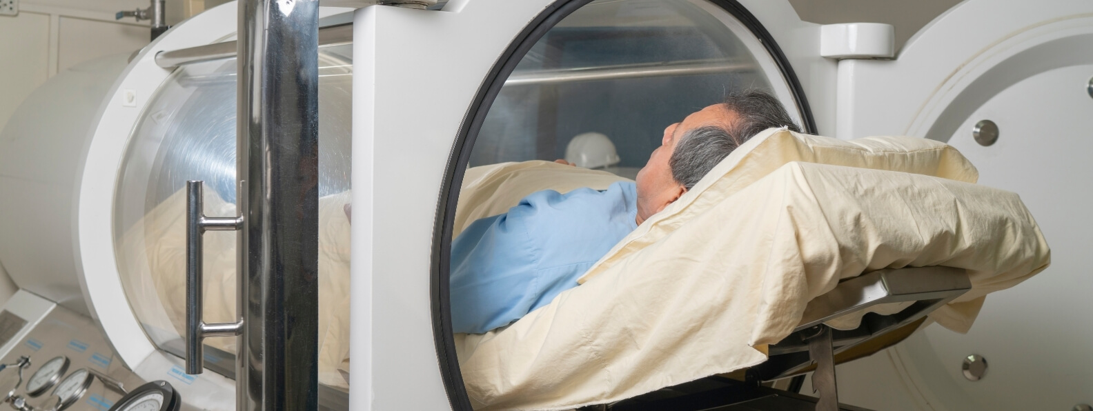
Hyperbaric oxygen therapy, commonly abbreviated as HBOT, may be used as an adjunct in selected traumatic injuries where tissue remains viable but oxygen delivery is critically impaired. The Undersea and Hyperbaric Medical Society recognizes crush injury, compartment syndrome, and other acute traumatic ischemias as hyperbaric indications. Medicare also identifies acute traumatic peripheral ischemia and crush injuries involving severed or replanted limbs among its covered conditions. (UHMS)
HBOT is not a general treatment for every fracture, contusion, surgical wound, or traumatic brain injury. Its most defensible role in acute trauma involves a defined threat to tissue perfusion, oxygenation, or reconstruction that persists after emergency surgical and vascular priorities have been addressed.
A high-energy injury can directly disrupt cells, capillaries, arteries, veins, lymphatic vessels, and supporting connective tissue. The resulting inflammatory response increases vascular permeability, allowing fluid to accumulate within and around the injured area.
As edema increases, the distance between capillaries and cells becomes greater. Pressure within confined tissue spaces may rise, while damaged or compressed vessels deliver less blood. Reduced perfusion produces additional hypoxia, which further damages capillary membranes and promotes more edema.
This creates a self-perpetuating cycle:
UHMS describes this edema-ischemia cycle as a central feature of crush injury, compartment syndrome, and other acute traumatic ischemias. HBOT is intended to interrupt the cycle by increasing oxygen delivery to viable tissue while reducing edema through hyperoxic vasoconstriction. (UHMS)
The clinical target is the zone of threatened tissue between clearly viable and irreversibly necrotic structures. HBOT cannot revive dead tissue and cannot overcome the complete absence of blood flow. It may help preserve marginal tissue that retains enough circulation to receive oxygen-rich plasma.
HBOT should never delay hemorrhage control, airway management, resuscitation, vascular repair, fracture stabilization, fasciotomy, debridement, or another time-critical operation.
Initial management may require:
HBOT is most useful when integrated into a coordinated trauma plan. It should support definitive care rather than compete with it.
A patient should not remain in a chamber while progressive ischemia requires an operation. Similarly, transfer to a distant hyperbaric center should not postpone vascular repair, fasciotomy, or debridement that can be performed immediately at the current hospital.
A crush injury occurs when a body part is compressed by a heavy object, machinery, structural collapse, vehicle, or another high-energy mechanism. The visible wound may underestimate the amount of deeper muscle and microvascular damage.
Crushed muscle can become ischemic and necrotic. As damaged muscle cells break down, they may release potassium, myoglobin, phosphate, and other intracellular substances into the circulation. Severe crush syndrome can contribute to dysrhythmia, shock, metabolic acidosis, acute kidney injury, and multiorgan failure. (PubMed Central (PMC))
Local treatment may involve debridement, fracture management, vascular reconstruction, wound coverage, and repeated assessment of tissue viability. Systemic care may require aggressive monitoring of electrolytes, renal function, urine output, acid-base status, and cardiovascular stability.
HBOT may be considered when viable muscle and soft tissue remain at risk because of severe edema, impaired microcirculation, or acute traumatic ischemia. Treatment does not replace removal of necrotic tissue or management of systemic crush syndrome.
A randomized, double-blind trial involving 36 patients with severe limb crush injuries compared surgery plus HBOT with surgery plus a sham exposure. Patients entered treatment within 24 hours after surgery and received two sessions daily for six days. The study reported improved wound healing and fewer repeat operations in the HBOT group, although its small sample limits the certainty and generalizability of the findings. (PubMed)
Acute compartment syndrome develops when pressure rises within a closed muscle compartment and compromises tissue perfusion. Fractures, crush injury, bleeding, reperfusion, tight casts or dressings, and extensive soft-tissue trauma can all contribute.
Possible warning findings include:
Pulse loss is a late and unreliable finding because arterial flow may continue after the microcirculation and muscle perfusion have become critically impaired.
Diagnosis depends on repeated clinical examination and, in selected patients, compartment-pressure measurement. Once acute compartment syndrome is diagnosed, urgent fasciotomy is the definitive treatment. AAOS guidance identifies acute compartment syndrome as a surgical emergency, and evidence shows that delayed decompression increases the risk of necrosis, infection, contracture, neurologic injury, and amputation. (PubMed)
HBOT must not be used to avoid or delay fasciotomy. Its possible roles are adjunctive:
If pressure continues to rise or clinical findings deteriorate, the patient needs surgical reassessment rather than another chamber session.
Open fractures with extensive soft-tissue injury are at risk for tissue necrosis, infection, nonunion, osteomyelitis, repeat reconstruction, chronic pain, and long-term disability.
HBOT has been studied most carefully in severe open fractures of the tibia. The international Hyperbaric Oxygen for Lower Limb Trauma, or HOLLT, trial enrolled 120 patients with severe open tibial fractures and compared standard trauma care with standard care plus 12 HBOT sessions.
The combined primary endpoint of tissue necrosis or infection within 14 days occurred in 43 percent of HBOT-assigned patients and 58 percent of controls. This difference did not reach statistical significance. Tissue necrosis considered separately occurred in 29 percent of HBOT patients and 53 percent of controls. The HBOT group also had fewer late complications and better functional outcomes during follow-up. (ResearchGate)
These findings are encouraging, but they require careful interpretation. The trial did not demonstrate a statistically significant reduction in its combined primary endpoint. It did show meaningful reductions in tissue necrosis and improvements in several longer-term outcomes.
A 2024 systematic review of HBOT in severe lower-limb soft-tissue trauma concluded that adjunctive treatment may improve wound healing and selected clinical outcomes. The authors also emphasized that the available literature remains limited by small study populations, variable protocols, and differences in injury severity and standard care. (Springer Link)
Current evidence supports considering HBOT for selected high-energy lower-limb injuries at experienced trauma centers. It does not justify routine treatment for every open fracture.
Traumatic peripheral ischemia occurs when an injury reduces blood flow to an extremity or other peripheral tissue. Causes may include arterial disruption, thrombosis, compression, extensive edema, vessel spasm, venous congestion, or microvascular destruction.
The first question is whether the blood supply can be restored directly. Vascular repair, thrombectomy, bypass, reduction of a dislocation, removal of external pressure, or correction of a constricting dressing may be required.
HBOT cannot substitute for revascularization. New oxygen cannot reach tissue when there is no functioning inflow. Its potential value begins after macroscopic circulation has been restored or when enough collateral and microvascular flow remains to deliver dissolved plasma oxygen.
HBOT may then support tissue by:
The goal is not merely to improve the appearance of the extremity. It is to preserve muscle, nerve, skin, bone, function, and the options available for reconstruction.
Replantation or revascularization of an amputated or nearly amputated body part can restore major arterial inflow while leaving the tissue vulnerable to edema, venous congestion, microvascular thrombosis, and reperfusion injury.
The surgical team must first establish technically adequate circulation. A thrombosed arterial anastomosis, obstructed vein, twisted pedicle, or compressive hematoma requires immediate surgical correction.
HBOT may be considered when the replanted tissue remains compromised after correctable mechanical causes have been addressed. Medicare’s national coverage determination specifically includes crush injuries and the suturing of severed limbs among covered hyperbaric indications. (Centers for Medicare & Medicaid Services)
The hyperbaric team should monitor:
HBOT may help preserve marginal tissue and reduce the level or extent of later debridement. It cannot guarantee replantation survival, and it should not delay revision of a failing vascular repair.
Major trauma frequently requires skin grafts, local flaps, pedicled flaps, or free-tissue transfer to cover exposed bone, vessels, tendons, nerves, or hardware.
HBOT is not recommended for a healthy graft or flap that is healing normally. It may be used when a reconstruction becomes compromised by hypoxia, ischemia, venous congestion, edema, or reperfusion injury.
Possible warning findings include:
Suspected flap compromise is first a surgical emergency. The reconstructive team must evaluate for thrombosis, hematoma, pedicle kinking, compression, tension, or technical failure.
UHMS notes that early initiation of HBOT after graft or flap compromise is identified may maximize the amount of tissue that remains viable. Treatment may reduce the need for complete regrafting or repeat reconstruction when enough circulation remains to support salvage. (UHMS)
During HBOT, the patient breathes oxygen while exposed to increased atmospheric pressure. This markedly raises arterial oxygen tension and increases the amount of oxygen dissolved directly in plasma.
The oxygen-rich plasma can move through functioning vessels and diffuse into injured tissue, including areas where red blood cell passage is limited by edema or microvascular narrowing.
At approximately 2 atmospheres absolute, hyperbaric exposure can substantially increase total blood oxygen content and create a stronger diffusion gradient into hypoxic tissue. UHMS emphasizes the combined ability to increase tissue oxygenation while reducing edema as a key rationale in acute traumatic ischemia. (UHMS)
Restoring blood flow is essential, but reperfusion itself can trigger oxidative stress, endothelial dysfunction, leukocyte activation, capillary leakage, and further edema.
This does not mean reperfusion should be avoided. It means that a successfully repaired vessel does not immediately normalize the entire microcirculation.
HBOT may influence this secondary phase by altering leukocyte-endothelial interactions, supporting tissue oxygenation, and limiting edema. The treatment is therefore most biologically plausible when delivered early, while the injured tissue remains viable and before secondary microvascular damage becomes irreversible.
The available clinical evidence does not identify one exact time limit that applies to every injury. Earlier consultation is generally preferable because salvage potential declines as necrosis becomes established. Reviews of traumatic ischemia recommend considering adjunctive HBOT as soon as practical after diagnosis and definitive stabilization. (PubMed)
The presence of trauma alone is not sufficient. A hyperbaric consultation should identify a specific oxygen-responsive problem and a defined treatment objective.
Potential candidates include patients with:
Factors supporting treatment may include documented perfusion compromise, severe swelling, evolving tissue discoloration, extensive muscle injury, high risk of necrosis, or a clinically salvageable reconstruction.
HBOT is less likely to help when:
Selection should be multidisciplinary, ideally involving trauma surgery, orthopedics, vascular surgery, plastic surgery, critical care, wound care, and hyperbaric medicine.
Hyperbaric protocols vary with injury severity, timing, chamber capability, and tissue response.
Commonly described regimens for acute traumatic ischemia use pressures around 2.0 to 2.5 atmospheres absolute for approximately 90 to 120 minutes. Treatments may be delivered more than once daily during the early period of greatest tissue threat, followed by a reduced frequency as edema and ischemia stabilize. (NCBI)
A typical course is not predetermined solely by diagnosis. It may be modified according to:
HBOT should be coordinated around the operating-room schedule. An indicated operation takes priority over a scheduled chamber session.
Treatment may be stopped when the tissue has stabilized, the clinical objective has been achieved, no further benefit is occurring, or the wound has progressed to irreversible necrosis requiring definitive surgery.
Traumatic wounds can change quickly, so serial assessment is essential.
The treatment team may monitor:
Photographs and standardized wound documentation can help demonstrate whether tissue is stabilizing. Perfusion imaging, transcutaneous oxygen measurement, angiography, or other vascular studies may be used when clinically appropriate, but no single test replaces bedside and operative assessment.
A temporary improvement in color during oxygen exposure does not by itself prove durable tissue survival. The meaningful outcomes are reduced necrosis, preservation of function, successful reconstruction, fewer operations, wound closure, and limb salvage.
Some patients considered for HBOT have multiple injuries, mechanical ventilation, invasive monitoring, chest tubes, infusions, or recent surgery.
Treatment requires a hospital-based hyperbaric program capable of maintaining the necessary level of care. Planning may involve:
Gas-filled spaces and devices may behave differently as pressure changes. Equipment must be specifically evaluated for chamber use.
A patient should not be placed in a chamber simply because HBOT might help the limb. The team must be able to protect the entire patient throughout transport, compression, treatment pressure, and decompression.
Potential complications of HBOT include:
An untreated pneumothorax is a major contraindication because trapped pleural gas may expand during decompression. Trauma patients with chest injury require particular attention before pressurization.
The operational risks may be as important as the direct treatment risks. Transporting an unstable patient away from the operating room or intensive care unit can interrupt resuscitation, delay surgery, or complicate access during deterioration.
HBOT should only be used when the expected tissue-salvage benefit exceeds these risks and the treatment can be delivered without weakening standard trauma care.
Clinical evidence is strongest for selected severe limb injuries, but it remains more limited than the evidence for many routine trauma interventions.
The existing evidence includes:
The evidence does not support claims that HBOT should be used routinely after uncomplicated fractures, minor soft-tissue injuries, or ordinary orthopedic surgery. It also does not establish HBOT as standard treatment for acute traumatic brain injury outside a research protocol.
The most appropriate interpretation is selective and time sensitive. HBOT may improve tissue preservation and functional recovery when acute trauma has created a reversible hypoxic or ischemic state that persists despite definitive surgical and vascular care.
A hospital seeking to use HBOT in acute trauma should establish referral and treatment pathways before the emergency occurs.
The pathway should define:
Early communication is particularly important. A hyperbaric team contacted only after extensive necrosis has developed may have little viable tissue left to salvage.
Hyperbaric therapy in acute trauma is best understood as a tissue-preservation strategy. It may increase oxygen delivery, reduce edema, and limit secondary ischemic injury, but only within a comprehensive system that provides rapid resuscitation, surgery, vascular care, infection prevention, reconstruction, and rehabilitation.
The chamber supports the trauma plan. It does not replace it.
A Practical Framework for Using Hyperbaric Oxygen in Tissue Salvage, Reconstruction, Trauma, Infection, and Radiation-Damaged Wounds Hyperbaric oxygen therapy, commonly
A Hospital Blueprint for Coordinated Diagnosis, Limb Preservation, Advanced Treatment, Hyperbaric Medicine, and Measurable Outcomes A multidisciplinary wound care program