How Hyperbaric Oxygen Therapy May Support Healing in Tissues Injured Months or Years After Radiation Treatment
Radiation therapy plays a critical role in the treatment of many cancers. Modern planning techniques allow clinicians to target tumors more precisely, but nearby healthy tissue may still receive enough radiation to produce long-term changes.

Some radiation effects occur during treatment and resolve afterward. Late radiation complications are different. They may develop months or years after radiation has ended and can become progressively more difficult to manage. Depending on the treatment field, patients may experience bleeding, pain, fibrosis, ulceration, tissue breakdown, impaired organ function, or bone exposure.
Hyperbaric oxygen therapy, commonly abbreviated as HBOT, may be considered for selected forms of delayed radiation injury. The Undersea and Hyperbaric Medical Society recognizes delayed radiation injury involving soft tissue and bone as an accepted indication for HBOT. Medicare also covers HBOT as an adjunct to conventional treatment for soft tissue radionecrosis and osteoradionecrosis. (UHMS)
HBOT is not appropriate for every symptom that develops after cancer treatment. Its clinical role depends on the affected tissue, severity of injury, exclusion of recurrent malignancy, previous treatments, and whether impaired vascularity is contributing meaningfully to the problem.
What Are Late Radiation Complications?
Late radiation tissue injury, sometimes called delayed radiation injury or radiation necrosis, refers to tissue damage that becomes clinically apparent after the acute effects of radiation have resolved.
Radiation can injure the endothelial cells lining small blood vessels. Over time, affected vessels may narrow, thrombose, or disappear. The surrounding tissue becomes less vascular, less cellular, and chronically hypoxic. Fibrosis may increase while the tissue’s ability to remodel, resist infection, and heal after minor trauma declines. (PubMed Central (PMC))
This process may remain clinically silent until an additional stress exposes the limited reserve of the irradiated tissue. Symptoms can begin after dental extraction, surgery, infection, pressure, instrumentation, or a minor wound. In other patients, tissue breakdown develops without an obvious triggering event.
Late radiation injury can affect:
- The bladder
- The rectum and lower bowel
- The jaw and other bones
- Oral and pharyngeal tissues
- The larynx
- The skin and chest wall
- Breast and axillary tissues
- Pelvic and gynecologic tissues
- Surgical wounds located within a previous radiation field
The symptoms depend on the organ involved, but the underlying tissue frequently shares a pattern of vascular damage, hypoxia, fibrosis, and reduced healing capacity.
Why Radiation Injury Can Progress Years Later
Radiation treatment may eliminate or control cancer while also initiating long-term changes in nearby normal tissue. Unlike an acute wound that moves through a predictable healing process, irradiated tissue may undergo continuing vascular and fibrotic remodeling.
As small-vessel density decreases, less oxygen reaches the tissue. Fibroblasts and other repair cells may function less effectively, while collagen becomes increasingly dense and disorganized. The tissue may become stiff, fragile, painful, and susceptible to breakdown.
A patient may appear fully recovered from cancer treatment and then develop hematuria, rectal bleeding, exposed jawbone, a nonhealing wound, or increasing fibrosis years later. The delayed timing can be confusing and distressing, particularly when the symptoms raise concern about cancer recurrence.
Late symptoms should never be attributed automatically to radiation history. Recurrent malignancy, infection, vascular disease, medication effects, and unrelated conditions must be considered before a diagnosis of delayed radiation injury is established.
Soft Tissue Radionecrosis
Soft tissue radionecrosis describes delayed radiation damage involving tissue other than bone. It may affect mucosa, skin, muscle, connective tissue, blood vessels, or internal organs.
Possible presentations include:
- Chronic ulceration or tissue breakdown
- Recurrent bleeding
- Pain and tissue induration
- Poor healing after surgery or instrumentation
- Fibrosis and restricted movement
- Fistula formation
- Recurrent infection
- Necrosis of skin, muscle, or mucosal tissue
Radiation cystitis and radiation proctitis are organ-specific forms of delayed soft tissue radiation injury. Other examples include laryngeal radionecrosis, chest-wall injury, vaginal or pelvic soft tissue necrosis, and nonhealing wounds within a previously irradiated field.
The diagnosis requires more than confirming that a tissue was exposed to radiation. Clinicians must evaluate the location and dose of the original radiation, the timing of symptoms, physical and imaging findings, infection status, and the possibility of persistent or recurrent cancer.
Osteoradionecrosis and Radiation-Damaged Bone
Osteoradionecrosis occurs when irradiated bone becomes devitalized and fails to heal adequately. It is most commonly discussed in the jaw after treatment for head and neck cancer, but radiation-associated bone injury may occur at other sites.
Patients may develop:
- Exposed bone
- Persistent oral or facial pain
- Drainage or fistula formation
- Infection
- Pathologic fracture
- Difficulty chewing or swallowing
- Trismus
- Failure of a dental extraction site to heal
Management may include oral hygiene measures, antimicrobial therapy when infection is present, limited debridement, medical antifibrotic regimens, resection, and reconstructive surgery. HBOT has historically been incorporated into some preventive and treatment protocols.
The role of HBOT in jaw osteoradionecrosis is now more controversial than its broader recognition as a hyperbaric indication might suggest. A 2024 joint guideline from the International Society of Oral Oncology, the Multinational Association of Supportive Care in Cancer, and the American Society of Clinical Oncology concluded that evidence supporting HBOT for the prevention or management of jaw osteoradionecrosis remains limited. (ASCO Publications)
This does not mean HBOT has no role in every case. It means treatment should not be applied automatically based on an older protocol alone. Decisions should consider the stage of disease, planned surgery, available reconstructive options, previous medical management, and the experience of the multidisciplinary team.
How HBOT Affects Irradiated Tissue
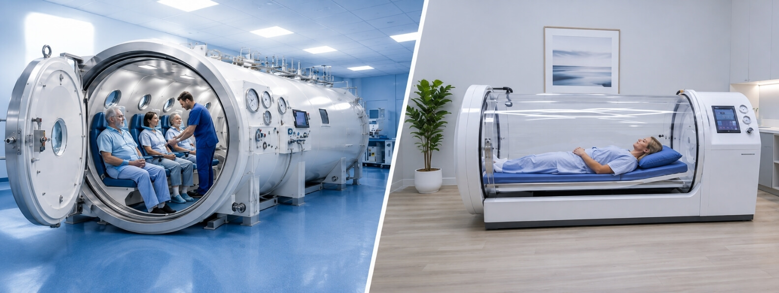
During HBOT, the patient breathes oxygen while exposed to increased atmospheric pressure inside a medical hyperbaric chamber. The increased oxygen partial pressure substantially raises the amount of oxygen dissolved in plasma.
This oxygen-rich plasma can move through functioning vessels at the edge of the radiation field and create a stronger diffusion gradient into hypoxic tissue. Each exposure temporarily increases tissue oxygen tension, but the objective of a full treatment course is broader than temporary hyperoxygenation.
Repeated treatments may support:
- Angiogenic signaling and new capillary formation
- Improved oxygen delivery between treatments
- Fibroblast activity and collagen remodeling
- Mucosal and epithelial repair
- Improved leukocyte function
- Modulation of chronic inflammation
- Healing after surgical debridement or reconstruction
HBOT cannot restore tissue that has already been completely destroyed. It also cannot remove necrotic bone, correct a fixed obstruction, close every fistula, or treat recurrent cancer. Its purpose is to improve the biologic environment of viable but compromised tissue and complement the necessary medical or surgical treatment.
A 2023 Cochrane review found evidence that HBOT may improve outcomes in selected late radiation injuries, including some injuries involving the bladder, rectum, bone, and head and neck tissues. The review also emphasized that many studies were small, clinically varied, or at risk of bias, leaving uncertainty about which patients benefit most and which protocols are optimal. (PubMed Central (PMC))
Radiation Cystitis and Bladder Injury
Chronic radiation cystitis may cause urinary frequency, urgency, pain, incontinence, reduced bladder capacity, and recurrent hematuria. Severe bleeding can produce clot retention, anemia, transfusion requirements, and repeated hospitalization.
HBOT is intended to improve the vascular condition of the bladder wall rather than simply stop one visible bleeding vessel. It is commonly considered after urologic evaluation has excluded recurrent malignancy and other causes of hematuria.
The strongest randomized evidence for HBOT in late pelvic radiation injury comes from radiation cystitis. The RICH-ART trial found improvement in patient-reported urinary symptoms after HBOT, and five-year follow-up published in 2025 reported that meaningful improvements were sustained in many initial responders. Some patients required another treatment course after symptoms recurred. (PubMed)
Patients with active clot retention, major blood loss, or urinary obstruction still require immediate urologic stabilization. HBOT is a restorative treatment, not a substitute for catheterization, clot evacuation, transfusion, cystoscopy, or other urgent interventions.
Radiation Proctitis and Bowel Injury
Chronic radiation proctitis may cause rectal bleeding, urgency, tenesmus, mucus discharge, pain, diarrhea, or fecal incontinence. Telangiectatic vessels in the irradiated rectal lining can rupture repeatedly, while fibrosis may reduce tissue compliance.
HBOT may be considered when symptoms persist after medical or endoscopic care, particularly when disease is diffuse, ulcerative, or difficult to treat without causing additional tissue injury.
The evidence is mixed. Some randomized and observational studies have reported improvement in bleeding and other symptoms, while another sham-controlled trial did not demonstrate a significant benefit for a broader group of chronic bowel symptoms. The 2023 Cochrane review concluded that HBOT may help selected patients but that certainty remains limited by differences in study populations and outcomes. (PubMed Central (PMC))
This variation reinforces the need to define the treatment target clearly. A patient with confirmed hemorrhagic radiation proctopathy is clinically different from a patient with nonspecific bowel symptoms caused by altered motility, malabsorption, pelvic floor dysfunction, or another gastrointestinal disorder.
Breast, Chest Wall, and Soft Tissue Fibrosis
Late radiation effects after breast cancer treatment may include pain, edema, skin changes, fibrosis, restricted shoulder movement, and tissue tightness. These symptoms can interfere with clothing, exercise, sleep, work, and daily activities.
A 2024 randomized clinical trial evaluated HBOT for late local toxic effects after breast irradiation. In the intention-to-treat analysis, being offered HBOT was associated with reduced fibrosis but not a statistically significant reduction in pain. Among participants who completed HBOT, both pain and fibrosis improved, although treatment uptake was low and the trial design requires careful interpretation. (JAMA Network)
These findings are promising but do not establish HBOT as routine treatment for all post-radiation breast symptoms. Lymphedema, shoulder dysfunction, recurrent disease, neuropathic pain, and musculoskeletal conditions should also be assessed and treated appropriately.
When a Hyperbaric Consultation May Be Appropriate
A hyperbaric evaluation may be reasonable when a patient has a documented history of therapeutic radiation and a clinically significant tissue complication consistent with delayed radiation injury.
Potential reasons for referral include:
- Persistent bladder or rectal bleeding
- Chronic radiation-associated ulceration
- Nonhealing wounds in an irradiated field
- Soft tissue necrosis
- Selected cases of osteoradionecrosis
- Tissue breakdown after surgery or dental treatment
- Radiation-associated pain and fibrosis in selected settings
- Planned reconstruction involving compromised irradiated tissue
- Failure of appropriate conventional treatment
The consultation should answer several questions:
- Is the current problem truly caused by delayed radiation injury?
- Has recurrent malignancy been evaluated appropriately?
- Is infection present?
- Is surgery, endoscopy, vascular care, or another intervention more urgent?
- Is the affected tissue viable enough to respond?
- What functional outcome is the treatment intended to improve?
- Can the patient complete a prolonged treatment schedule safely?
HBOT should be integrated into a defined clinical plan rather than added because symptoms have been difficult to treat.
What an HBOT Course May Involve
Treatment protocols vary by anatomy, severity, prior care, and whether surgery is planned. Many delayed radiation injury protocols involve treatment five days per week for several weeks.
A course often includes approximately 30 to 40 sessions, although additional treatments may be considered when the patient is improving or when HBOT is coordinated with surgery. Treatment pressures commonly fall between approximately 2.0 and 2.5 atmospheres absolute, with prescribed oxygen periods and air breaks determined by the hyperbaric physician. Clinical trials and systematic reviews have used varying pressures, durations, and treatment numbers, and no single schedule applies to every radiation complication. (PubMed Central (PMC))
Clinical response should be evaluated throughout treatment. Measures may include:
- Frequency and severity of bleeding
- Wound dimensions and tissue quality
- Pain and medication requirements
- Urinary or bowel function
- Range of motion
- Need for transfusion or hospitalization
- Progress toward surgery or reconstruction
- Patient-reported quality of life
Improvement may occur during the treatment course or continue afterward as vascular and tissue remodeling progresses.
HBOT Remains an Adjunct to Multidisciplinary Care
Late radiation complications often require more than one specialty. Depending on the tissue involved, care may include radiation oncology, urology, colorectal surgery, gastroenterology, oral and maxillofacial surgery, otolaryngology, plastic surgery, wound care, physical therapy, and hyperbaric medicine.
Conventional treatment may include:
- Endoscopic control of bleeding
- Debridement or removal of necrotic tissue
- Antimicrobial therapy
- Dental or oral surgical care
- Medical treatment of fibrosis
- Wound dressings and reconstructive surgery
- Bladder irrigation or clot evacuation
- Nutritional and anemia management
- Pelvic floor or physical rehabilitation
CMS coverage language specifically describes HBOT for soft tissue radionecrosis and osteoradionecrosis as adjunctive treatment. This reflects the clinical reality that oxygen therapy is most useful when paired with appropriate management of the structural, infectious, oncologic, and functional components of the injury. (Centers for Medicare & Medicaid Services)
Risks and Practical Considerations
HBOT is generally well tolerated in appropriately screened patients, but it carries recognized risks. These include middle-ear or sinus barotrauma, temporary myopic vision changes, claustrophobia, blood glucose changes, pulmonary oxygen effects, and rare oxygen-induced seizure.
An untreated pneumothorax is generally considered an absolute contraindication. Pulmonary disease, difficulty equalizing ear pressure, unstable cardiac disease, seizure risk, implanted medical devices, and medication concerns require individualized assessment.
The treatment schedule can also be burdensome. Patients may attend daily sessions for six to eight weeks while continuing cancer surveillance, specialist visits, wound care, or other treatments. Transportation, work, caregiving responsibilities, fatigue, urinary urgency, bowel symptoms, and mobility limitations can affect whether the plan is realistic.
These considerations should be discussed before treatment begins. A technically appropriate therapy still needs to be practical, safe, and connected to outcomes that matter to the patient.
Setting Realistic Expectations
HBOT should not be presented as a universal remedy for radiation damage. Evidence is stronger for certain conditions, such as chronic radiation cystitis, than for others. Research involving rectal injury, breast toxicity, skin necrosis, and head and neck complications is promising in some areas but inconsistent or limited in others. Jaw osteoradionecrosis remains a particularly important area of clinical debate. (PubMed)
Treatment success may mean complete resolution of bleeding or wound closure, but it can also mean fewer procedures, improved tissue quality, reduced pain, greater function, or a safer reconstructive operation.
For patients who believed their cancer treatment was behind them, the appearance of a delayed complication can feel like an unexpected return to illness. Compassionate care requires acknowledging that burden while providing a careful, evidence-based explanation of what HBOT may and may not accomplish.
When the diagnosis is clear, recurrent cancer has been appropriately evaluated, and chronic tissue hypoxia is contributing to the problem, HBOT may provide a meaningful opportunity to improve the healing capacity of previously irradiated tissue.