

Integrating HBOT into Surgical Practice
A Practical Framework for Using Hyperbaric Oxygen in Tissue Salvage, Reconstruction, Trauma, Infection, and Radiation-Damaged Wounds Hyperbaric oxygen therapy, commonly
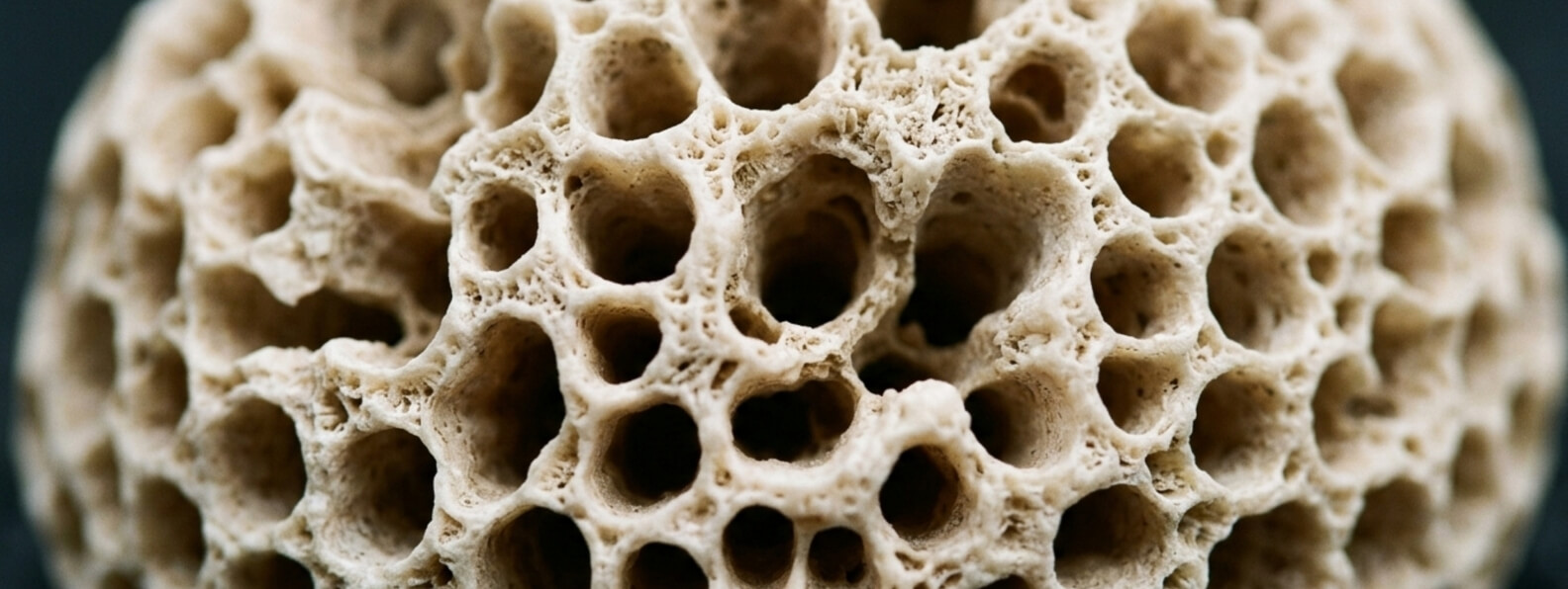
Osteomyelitis is an infection involving bone or bone marrow. It may develop through bloodstream spread, direct contamination after trauma or surgery, extension from a nearby wound, or infection associated with orthopedic hardware. Treatment can be difficult because infected bone may contain poorly perfused tissue, necrotic fragments, bacterial biofilm, and structural areas that systemic antibiotics cannot easily reach.
Most cases of osteomyelitis are treated with antimicrobial therapy, surgical source control, or a combination of both. Hyperbaric oxygen therapy, commonly abbreviated as HBOT, is not a first-line treatment for uncomplicated bone infection. Its recognized role is primarily as an adjunct for chronic refractory osteomyelitis that persists or recurs despite appropriate medical and surgical management. Medicare coverage similarly identifies chronic refractory osteomyelitis that has not responded to conventional treatment as a covered HBOT condition. (UHMS)
Refractory osteomyelitis generally refers to a bone infection that persists or returns after appropriate treatment has been attempted. It may also describe an acute infection that continues to progress despite accepted management.
The term should not be applied simply because osteomyelitis requires several weeks of antibiotics. Bone infections commonly need prolonged treatment, and radiographic abnormalities may remain visible after the patient has begun to improve. Persistent imaging changes alone do not necessarily establish treatment failure.
A refractory infection is more likely to involve evidence such as:
Definitions and monitoring methods vary according to the anatomic site. For native vertebral osteomyelitis, the Infectious Diseases Society of America recommends interpreting symptoms, examination findings, inflammatory markers, microbiology, and imaging together. Persistent pain, neurologic deficits, elevated inflammatory markers, or abnormal imaging findings considered individually do not necessarily prove that treatment has failed. (Infectious Diseases Society of America)
Before labeling an infection refractory, the clinical team should determine whether the original diagnosis was correct, whether the causative organism was identified, whether the antimicrobial regimen was appropriate, and whether residual infected or necrotic tissue remains.
Healthy bone is living, vascular tissue that continually remodels. Osteomyelitis can disrupt that environment by damaging the microcirculation and separating devitalized bone from its blood supply.
A fragment of necrotic bone, known as a sequestrum, may serve as a protected surface for microorganisms. Antibiotics delivered through the bloodstream may have limited access to this nonviable tissue. Infections involving plates, screws, prosthetic material, or other implanted hardware may also develop biofilm, a structured microbial community that adheres to a surface and becomes more tolerant of antimicrobial therapy and host immune defenses.
Several clinical factors can further complicate treatment:
These factors do not automatically establish an indication for HBOT. They help explain why some infections remain active even after an apparently appropriate course of care.
During HBOT, the patient breathes oxygen while exposed to increased atmospheric pressure inside a medical hyperbaric chamber. The elevated oxygen partial pressure substantially increases the amount of oxygen dissolved in plasma and can raise oxygen tension in tissues supplied by functioning blood vessels.
In refractory osteomyelitis, several physiologic effects may be clinically relevant.
HBOT may improve oxygen availability in hypoxic bone and surrounding soft tissue. This matters because immune cells use oxygen-dependent mechanisms to kill certain microorganisms. Severe local hypoxia can impair this oxidative activity even when circulating leukocyte numbers are normal.
Controlled hyperoxia may also support:
These mechanisms are complementary rather than curative on their own. HBOT does not remove sequestra, drain an abscess, stabilize an infected fracture, restore flow through an obstructed artery, or eliminate a colonized implant. Reviews from the Undersea and Hyperbaric Medical Society emphasize that the best outcomes are generally obtained when HBOT is delivered alongside culture-directed antibiotics and appropriate surgical management. (PubMed)
When necrotic bone, purulent material, unstable tissue, or infected hardware is present, surgery often remains an essential part of treatment.
Depending on the location and severity of the infection, surgical management may include:
HBOT should not be used to postpone urgent source control. A patient with sepsis, progressive neurologic compromise, an undrained abscess, extensive necrosis, or mechanical instability requires prompt evaluation by the appropriate surgical and infectious-disease teams.
There are circumstances in which extensive surgery or hardware removal may carry substantial risk. Cranial, spinal, sternal, and pediatric osteomyelitis can present situations in which aggressive resection is technically difficult or potentially disabling. In selected cases, a coordinated trial of limited surgery, culture-directed antibiotics, and HBOT may be considered when the expected harm of more extensive intervention is high. (PubMed)
HBOT is not an antibiotic and should not be used as a substitute for microbiologic diagnosis or systemic antimicrobial therapy.
Whenever clinically feasible, deep tissue or bone cultures should be obtained to identify the causative organism and guide treatment. Superficial wound swabs may not accurately represent organisms within infected bone. The most useful specimen is generally obtained from bone or deep tissue using a technique that minimizes contamination.
Antibiotic selection depends on:
The required duration varies considerably. IDSA guidance for native vertebral osteomyelitis recommends approximately six weeks of parenteral or highly bioavailable oral antimicrobial therapy for most bacterial cases. For diabetes-related foot osteomyelitis, guideline recommendations distinguish between infections managed with bone resection or amputation and those treated without bone removal. These examples illustrate why treatment duration must be based on the infection site and clinical scenario rather than a universal osteomyelitis schedule. (Infectious Diseases Society of America)
A hyperbaric consultation may be reasonable when osteomyelitis has continued or recurred despite a coordinated treatment plan that includes appropriate antimicrobial therapy and surgical evaluation.
Potential candidates may include patients with:
The decision should be made by a multidisciplinary team whenever possible. The team may include orthopedic surgery, podiatric surgery, infectious disease, plastic surgery, vascular surgery, wound care, radiology, and hyperbaric medicine.
The presence of osteomyelitis alone is not enough. The team should identify why conventional treatment has failed and whether HBOT addresses a meaningful component of that failure.
Osteomyelitis associated with a diabetic foot ulcer deserves particular attention because HBOT may be considered under more than one clinical framework.
A patient may qualify for HBOT because of chronic refractory osteomyelitis. A different patient may qualify because of a sufficiently advanced diabetic lower-extremity wound that has not responded to standard wound care. These pathways overlap, but they are not identical.
The IWGDF/IDSA diabetic foot infection guideline advises against using HBOT solely for the purpose of treating a diabetic foot infection. This does not mean HBOT can never be used when a diabetic foot ulcer also contains infected bone. It means that HBOT should not replace surgical drainage, debridement, systemic antibiotics, vascular assessment, or other established infection treatment. (Infectious Diseases Society of America)
For a diabetic foot ulcer with osteomyelitis, the treatment plan should also address:
When infection and peripheral artery disease occur together, urgent surgical and vascular consultation may be required to coordinate drainage, debridement, and revascularization. (Infectious Diseases Society of America)
A hyperbaric evaluation should confirm both the clinical indication and the patient’s ability to tolerate treatment safely.
The medical review commonly includes:
MRI is frequently used to define the extent of osteomyelitis and associated soft-tissue disease, although postoperative changes and chronic abnormalities can complicate interpretation. Other imaging methods may be selected when MRI is contraindicated or when the clinical question requires a different modality. IDSA guidance for suspected vertebral osteomyelitis recommends MRI as the preferred initial imaging study and identifies nuclear imaging, CT, or PET as possible alternatives in selected circumstances. (Infectious Diseases Society of America)
The evaluation should also determine whether the patient is receiving adequate nutrition, antimicrobial therapy, pressure relief, vascular care, and wound management. HBOT is less likely to help when these foundational issues remain unresolved.
HBOT protocols for refractory osteomyelitis are individualized according to infection severity, anatomic location, surgical timing, comorbidities, and response.
A commonly described approach involves treatment once daily, five to seven days per week, at approximately 2.0 to 3.0 atmospheres absolute. Oxygen exposure commonly lasts 90 to 120 minutes, with air breaks incorporated when appropriate. A course may continue for approximately four to six weeks, although some patients require fewer or additional sessions. (PubMed)
HBOT may be started after debridement, delivered during an extended antimicrobial course, or coordinated around staged reconstructive procedures. There is no single protocol appropriate for every bone infection.
During treatment, clinicians should continue to evaluate:
A predetermined number of sessions should not prevent reassessment. Lack of meaningful progress should prompt the team to reconsider residual necrotic bone, inadequate source control, resistant organisms, biofilm, impaired blood flow, nonadherence, or an alternative diagnosis.
HBOT is generally well tolerated when patients are appropriately screened, but treatment is not risk free.
Potential complications include:
An untreated pneumothorax is generally considered an absolute contraindication. Significant pulmonary disease, inability to equalize ear pressure, unstable heart failure, seizure risk, medication interactions, and implanted devices require individualized evaluation.
The treatment schedule can also be demanding. A patient may need daily chamber sessions while receiving intravenous antibiotics, undergoing dressing changes, attending surgical appointments, and managing limited mobility. Transportation, caregiver support, employment obligations, and treatment fatigue should be considered before beginning a lengthy course.
These practical barriers are not secondary concerns. A clinically appropriate treatment can only be effective when the patient can participate in the complete care plan.
The evidence supporting HBOT for refractory osteomyelitis is based largely on physiologic studies, animal research, observational cohorts, case series, and systematic reviews of nonrandomized data.
No randomized clinical trials have directly established the effect of HBOT in refractory osteomyelitis. Published reports generally describe favorable outcomes when HBOT is added to surgery and culture-directed antimicrobial therapy, but differences in infection location, patient selection, treatment protocols, follow-up, and definitions of remission limit certainty. (PubMed)
More recent observational research continues to evaluate outcomes and factors associated with treatment failure, but it does not remove the need for controlled trials. HBOT should therefore be presented as an evidence-supported adjunct for selected refractory cases, not as a guaranteed cure or routine treatment for every bone infection. (PubMed)
Medicare recognizes chronic refractory osteomyelitis that is unresponsive to conventional medical and surgical management as a covered indication for chamber-based HBOT. Coverage requirements and documentation standards may vary among insurers and do not independently establish that treatment is appropriate for a particular patient. (Centers for Medicare & Medicaid Services)
The strongest clinical rationale exists when the infection remains active despite appropriate care, viable tissue can still be preserved, and increased tissue oxygenation can complement a clear surgical and antimicrobial strategy.
Hyperbaric therapy works best as part of a disciplined effort to control infection, preserve function, and restore healthy tissue. For patients facing repeated procedures, prolonged antibiotics, or the possibility of losing bone or limb function, that coordinated approach should remain clinically rigorous while recognizing the significant personal burden of living with a persistent bone infection.
A Practical Framework for Using Hyperbaric Oxygen in Tissue Salvage, Reconstruction, Trauma, Infection, and Radiation-Damaged Wounds Hyperbaric oxygen therapy, commonly
A Hospital Blueprint for Coordinated Diagnosis, Limb Preservation, Advanced Treatment, Hyperbaric Medicine, and Measurable Outcomes A multidisciplinary wound care program