

Integrating HBOT into Surgical Practice
A Practical Framework for Using Hyperbaric Oxygen in Tissue Salvage, Reconstruction, Trauma, Infection, and Radiation-Damaged Wounds Hyperbaric oxygen therapy, commonly
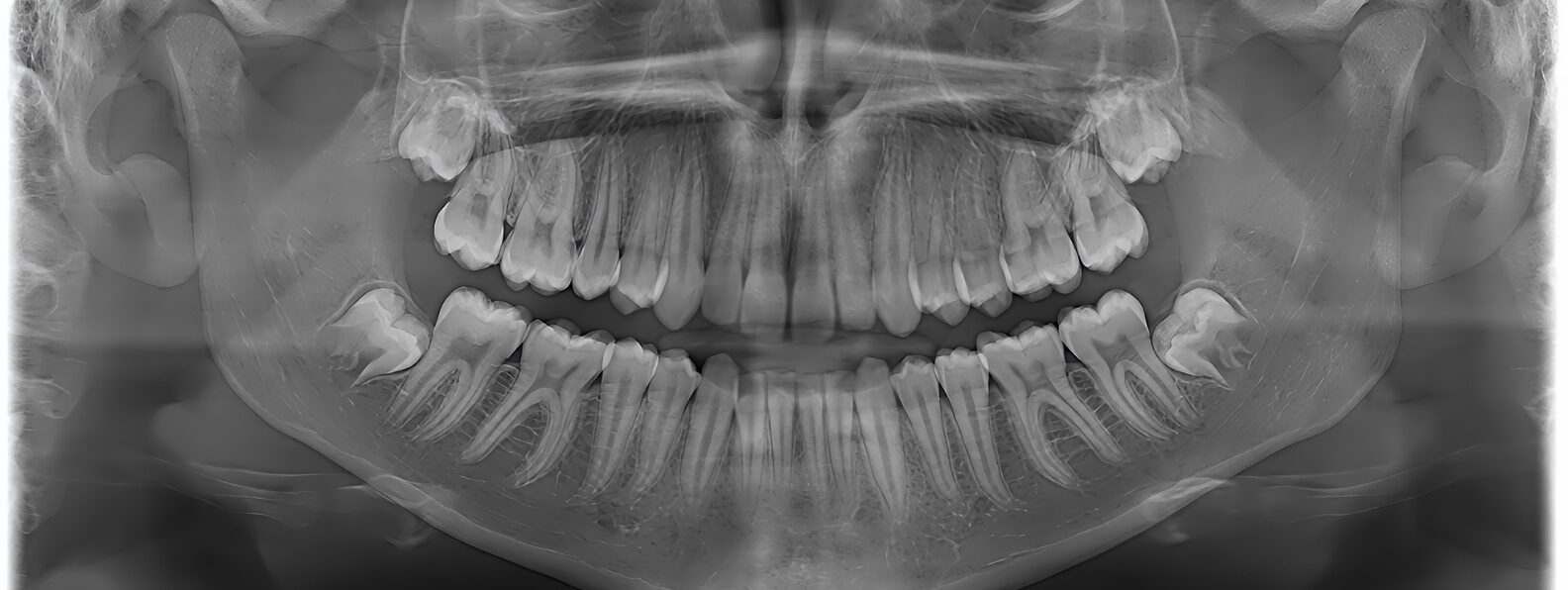
Dental surgery after head and neck radiation requires more planning than the same procedure in nonirradiated tissue. Radiation may reduce vascular density, alter bone remodeling, increase fibrosis, and weaken the ability of oral tissues to recover from trauma. An extraction socket or implant osteotomy that would ordinarily heal without difficulty can become a persistent wound within a heavily irradiated region.
The principal concern is osteoradionecrosis, commonly abbreviated as ORN. This condition involves devitalized irradiated bone and may present with exposed bone, pain, infection, drainage, fistula formation, impaired oral function, or pathologic fracture.
The goal is not to deny necessary dental care to cancer survivors. An infected or painful tooth can create its own significant risks. The clinical objective is to determine whether surgery is truly necessary, understand the radiation exposure at the proposed site, reduce avoidable tissue injury, and follow the patient until healing is clearly established.
Radiation can injure the endothelial cells lining the small blood vessels that supply bone and adjacent soft tissue. Over time, progressive vascular loss, fibrosis, and changes in cellular activity may leave the tissue less capable of responding to infection or surgical trauma.
Normal extraction healing depends on blood-clot formation, inflammatory cell activity, development of granulation tissue, mucosal coverage, and gradual remodeling of the socket. These processes require adequate vascularity, oxygen delivery, immune function, and viable bone.
Within an irradiated field, several parts of this sequence may be impaired:
The 2024 joint guideline from the International Society of Oral Oncology, Multinational Association of Supportive Care in Cancer, and American Society of Clinical Oncology considers jaw sites exposed to at least 50 Gy to be at increased risk for ORN. It also identifies dentoalveolar surgery, poor oral hygiene, and tobacco use as important risk factors. (OMS Group)
A history of head and neck radiation does not provide enough information to estimate surgical risk. The total cancer-treatment dose may differ substantially from the dose delivered to a specific extraction or implant site.
Before finalizing the dental treatment plan, the clinical team should review the original radiation plan whenever it is available. The review should identify:
Current guidance strongly recommends reviewing the dose delivered to the mandible and maxilla before planning dental treatment in a patient with previous head and neck radiation. (OMS Group)
The mandible is generally of particular concern because its anatomy and vascular supply may leave portions of the bone more vulnerable to radiation injury. Risk is still site specific. An anterior mandibular tooth outside the high-dose field may present a different clinical problem from a posterior mandibular molar located within a region that received a substantial dose.
Time since radiation should also be documented, but no elapsed interval makes previously irradiated bone completely equivalent to nonirradiated bone. ORN can develop years after treatment, particularly when surgery, infection, trauma, or severe dental disease challenges the tissue’s limited healing reserve. The history should therefore remain part of lifelong dental planning.
The decision to operate should begin with a diagnosis, not simply with the observation that a tooth has a poor appearance.
The examination may include:
Clinicians should look for evidence that ORN may already be present. Exposed bone, a nonhealing socket, a mucosal or cutaneous fistula, unexplained radiographic sclerosis or lysis, and bone that can be probed through a periodontal defect may indicate existing disease rather than uncomplicated dental pathology. The current guideline defines ORN using clinical and radiographic findings within an anatomic site previously exposed to therapeutic head and neck radiation.
When ORN is suspected, the patient may require evaluation by oral and maxillofacial surgery, head and neck surgery, dental oncology, or another experienced specialist before the tooth is removed. Recurrent malignancy must also remain in the differential diagnosis when findings are atypical, progressive, or accompanied by a suspicious mass.
Preserving a treatable tooth may avoid surgical injury to irradiated bone. Current guidance recommends offering alternatives to extraction for teeth in high-risk areas unless recurrent infection, uncontrolled pain, or another problem cannot be resolved conservatively. (OMS Group)
Depending on the tooth and clinical circumstances, alternatives may include:
Conservative treatment should not become indefinite postponement of necessary source control. A nonrestorable tooth with recurrent abscesses, cellulitis, uncontrolled pain, or progressive infection can threaten surrounding tissue and the patient’s general health.
The decision should balance two risks: the risk of surgical injury to irradiated bone and the risk of allowing active dental disease to continue.
When extraction is necessary, the procedure should be completed by a clinician who understands the patient’s radiation history and ORN risk. Referral to an oral and maxillofacial surgeon or dental oncologist is often appropriate for high-dose sites, complex teeth, previous healing problems, or patients with established radiation injury.
The operative objective is to remove the dental source of disease while producing as little additional injury as reasonably possible. The surgical plan may consider:
There is no single extraction technique proven to prevent ORN in every patient. Procedure design should reflect the tooth, anatomic site, radiation dose, soft-tissue condition, and the surgeon’s ability to achieve stable healing without creating additional trauma.
A painful or infected tooth should not be treated as an elective cosmetic issue. Patients often arrive after months of discomfort because they were told that extraction was impossible after radiation. In many cases, necessary surgery can still be performed, but it requires risk-adapted planning rather than a blanket refusal.
The 2024 ISOO-MASCC-ASCO guideline recommends considering oral antibiotics before and after invasive dental procedures in patients at higher risk because the treatment site lies within an irradiated portion of the mandible or maxilla. The evidence quality is low, so antibiotic selection and duration should be individualized rather than applied as one universal regimen. (OMS Group)
Antibiotics do not restore vascularity and should not be expected to compensate for retained infection, traumatic surgery, or inadequate follow-up. Their role is to reduce perioperative bacterial burden when the anticipated risk justifies their use.
For patients whose extraction site shows delayed healing, the guideline allows antiseptic rinses such as chlorhexidine or povidone-iodine to be used at least twice daily until adequate healing is confirmed. The patient should remain under close dental or surgical observation rather than relying on a rinse while exposed bone or infection progresses. (OMS Group)
Antimicrobial products should be selected with attention to allergies, local protocols, renal function, medication interactions, microbial findings, and the presence of active infection.
Pentoxifylline and tocopherol, commonly called the PENTO regimen, have been studied as a strategy for modifying radiation-associated fibrosis and supporting healing.
The 2024 guideline gives a weak recommendation for considering pentoxifylline 400 mg twice daily and tocopherol 1,000 IU once daily in cancer-free patients undergoing invasive dental procedures at sites that received at least 50 Gy. The suggested period is at least one week before the procedure and four weeks afterward, preferably until the socket has healed. (OMS Group)
This is not a universal protocol and should not be self-initiated. The evidence is based primarily on observational studies rather than randomized trials. Pentoxifylline and high-dose tocopherol may be inappropriate in patients with increased bleeding risk or certain renal, cardiac, hepatic, or medication-related concerns. The guideline specifically emphasizes evaluating contraindications before prescribing the regimen. (OMS Group)
The prescribing clinician should coordinate with the surgeon and the patient’s medical team, particularly when anticoagulants, antiplatelet agents, cardiovascular disease, renal impairment, or active cancer are present.
Dental implants can improve mastication, speech, prosthetic stability, and quality of life after head and neck cancer treatment. Implant placement also creates a surgical osteotomy within irradiated bone and therefore requires careful risk assessment.
Current guidance recommends avoiding implants in high-risk ORN zones unless less invasive methods of restoring oral function are not feasible. (OMS Group)
Implant planning should consider:
Implant survival and freedom from ORN are different outcomes. An implant may integrate successfully while the surrounding tissue remains vulnerable to later breakdown. Patients require long-term hygiene, prosthetic maintenance, and surveillance for peri-implant inflammation, exposed bone, pain, or drainage.
Hyperbaric oxygen therapy has historically been used before and after dental extraction in irradiated bone. Traditional protocols were intended to increase tissue oxygenation and stimulate angiogenesis before surgery.
Routine prophylactic HBOT is no longer recommended for every patient undergoing dental extraction after head and neck radiation. The 2024 ISOO-MASCC-ASCO guideline concluded that available evidence shows limited preventive benefit. It allows that HBOT may still be offered selectively when a substantial volume of the planned surgical site received more than 50 Gy. (OMS Group)
The HOPON randomized trial studied patients undergoing extraction or implant placement in a mandible exposed to more than 50 Gy. ORN occurred in 6.4 percent of the HBOT group and 5.7 percent of the control group, which did not demonstrate a routine preventive advantage from HBOT. All participants also received perioperative antibiotics and chlorhexidine. (PubMed)
A selective hyperbaric consultation may still be reasonable when the patient has several high-risk features, such as:
The expected benefit should be defined before treatment begins. HBOT should not be presented as a guarantee against ORN or used to compensate for untreated infection, incomplete surgery, or inadequate follow-up.
Follow-up should continue until mucosal closure and stable healing are clearly established. A patient should not be considered healed simply because pain has improved or the immediate postoperative period was uneventful.
Clinicians should monitor for:
Delayed healing should prompt reassessment of infection, retained bone or root fragments, radiation dose, oral hygiene, tobacco exposure, nutritional status, glucose control, prosthetic trauma, and possible ORN.
Current guidance recommends close clinical and radiographic surveillance when ORN is suspected or when abnormalities are detected.
Early specialist assessment is preferable to repeated short antibiotic courses without establishing why the socket remains open. Antibiotics may suppress a secondary infection while necrotic bone continues to progress underneath the mucosa.
Dental surgery in irradiated bone is safest when the dentist, oral surgeon, radiation oncologist, head and neck team, and hyperbaric physician communicate directly.
The radiation oncologist can provide site-specific dose information. The dental team can determine whether the tooth is salvageable. The surgeon can design the least disruptive effective procedure. The medical team can manage anticoagulation, diabetes, nutrition, tobacco dependence, and other systemic risks. A hyperbaric consultation can be added when the site and procedure create a credible need for adjunctive oxygen therapy.
For the patient, the plan should be understandable. They should know why surgery is necessary, how radiation affects healing, what steps are being taken to reduce risk, and which symptoms require prompt evaluation.
The presence of irradiated bone changes the treatment plan, but it does not eliminate the possibility of appropriate dental care. Careful dosimetric review, conservative treatment when feasible, risk-adapted surgery, and documented healing surveillance allow clinicians to address painful or infected teeth while reducing the likelihood of preventable complications.
A Practical Framework for Using Hyperbaric Oxygen in Tissue Salvage, Reconstruction, Trauma, Infection, and Radiation-Damaged Wounds Hyperbaric oxygen therapy, commonly
A Hospital Blueprint for Coordinated Diagnosis, Limb Preservation, Advanced Treatment, Hyperbaric Medicine, and Measurable Outcomes A multidisciplinary wound care program